
 E-mail Alert
 RSS
RSS
Preparation and optical characterization of large-area self-assembled gold nanoparticle superlattice films
-
摘要:
自组装贵金属纳米颗粒超晶格等离激元与光场的耦合能够激发极化激元模式,在增强光谱及传感等领域具有重要的应用前景。然而目前自组装方法制备超晶格薄膜层数较难控制,同时样品尺寸较小,限制了相关应用的发展。本文基于润湿增强的界面自组装方法可快速、大面积制备单层密排纳米颗粒薄膜的特性,采用逐层堆叠方法制备了具有不同层数的大面积、均匀分布的金纳米颗粒超晶格薄膜样品。实验及计算透/反射光谱表明,所制备超晶格样品能够有效激发极化激元模式,同时高阶极化激元模式随着超晶格层数的增加也可被有效激发。此外,通过调整纳米颗粒尺寸也可有效调制极化激元模式的共振峰位。这些研究为制备大面积高质量纳米颗粒超晶格薄膜提供了一种有效的方案,有望用于高性能微纳光子器件的设计。
Abstract:Self-assembly of noble metal nanoparticles into superlattices can couple with plasmonic modes and light fields, holding significant promise in enhanced spectroscopy and sensing applications. However, controlling the number of layers in these self-assembled superlattice films is challenging, and small sample sizes limit their potential applications. In this study, based on a wetting-enhanced interfacial self-assembly method, we demonstrate the rapid and large-scale fabrication of monolayer densely packed nanoparticle films. Additionally, a layer-by-layer stacking method is employed to fabricate large-area, uniformly distributed gold nanoparticle superlattice films with different numbers of layers. Both experimental and computational transmission/reflection spectra indicate that the prepared superlattice samples effectively excite plasmonic modes, and higher-order plasmonic modes can also be efficiently excited with increasing superlattice layers. Moreover, adjusting the nanoparticle size enables effective modulation of the resonance peak positions of plasmonic modes. These findings provide an effective approach for the large-scale fabrication of high-quality nanoparticle superlattice films, holding promise for the design of high-performance micro/nano photonic devices.
-
Overview: With the deepening development of nanotechnology, scientific research has increasingly focused on the unique photoelectric response properties of self-assembled noble metal nanoparticle superlattice structures. These structures effectively confine and enhance light fields, giving rise to a variety of plasmonic modes with significant implications for applications in spectroscopy enhancement, biosensing, and the development of nanophotonic devices. However, current self-assembly techniques face challenges in fabricating large-area, multilayer, and evenly distributed nanoparticle superlattice films, which limits their practical advancement. This study addresses this issue by introducing an innovative wetting-enhanced interfacial self-assembly method. This technique ensures rapid and uniform deposition over a large area while forming single-layer nanoparticle films. By employing a layer-by-layer stacking approach, the researchers successfully prepared a series of gold nanoparticle superlattice films with varying numbers of layers. Experimental measurements and theoretical computations of transmission/reflection spectra confirm that these films can effectively excite plasmonic modes. Furthermore, as the number of superlattice layers increases, higher-order plasmonic modes are also efficiently excited. Additionally, precise tuning of the gold nanoparticle size enables accurate control over the resonance peak positions of plasmonic modes. In summary, this study presents a novel approach for large-scale fabrication of high-quality multilayer gold nanoparticle superlattice films and reveals the critical role of nanoparticle size and superlattice layer count in governing plasmonic behavior. This research paves the way for the design and construction of advanced micro- and nanophotonic devices that leverage surface plasmon effects, thereby opening up new avenues in the field.
-

-
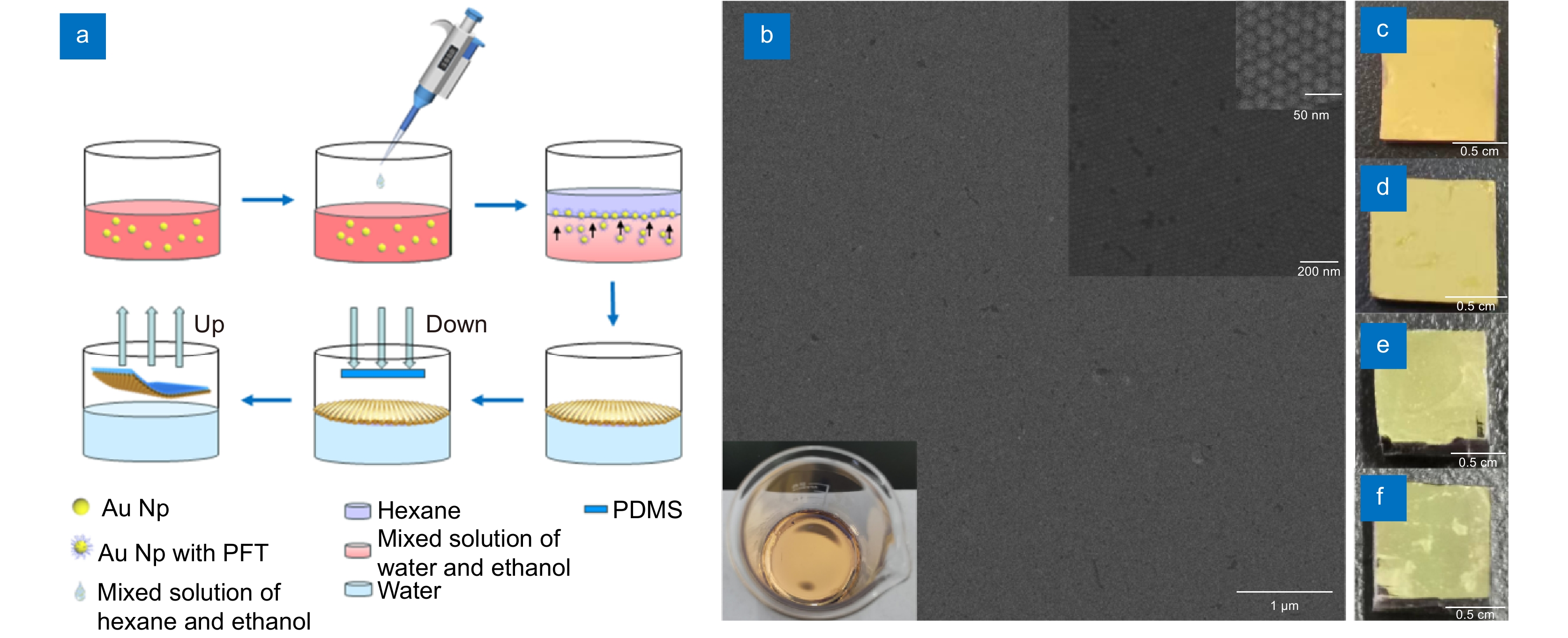
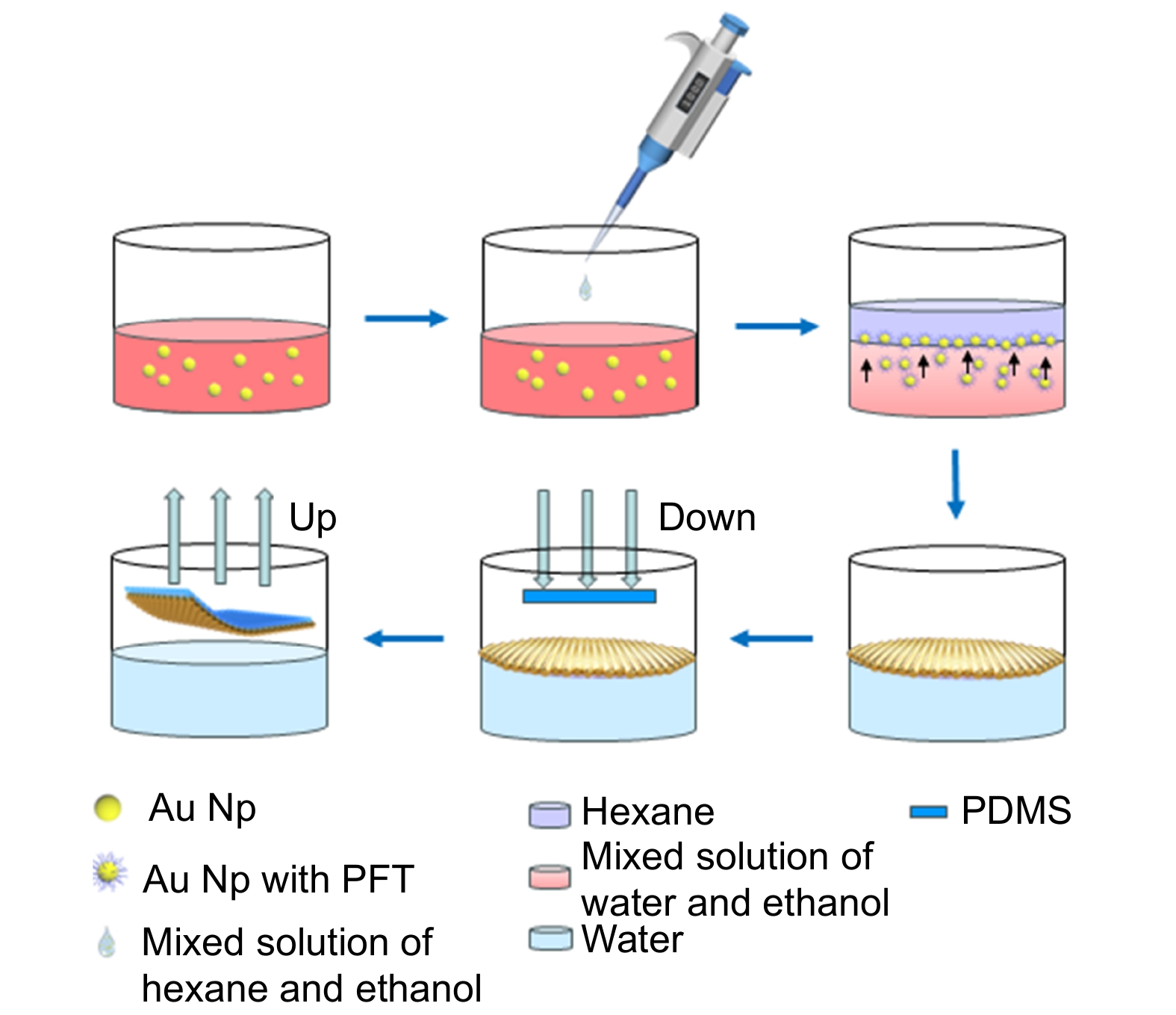
图 1 (a)由PFT诱导的金NPs在水-正己烷界面上的自组装以及薄膜转移过程示意图;(b)尺寸为20 nm,颗粒间间隔为1~2 nm的单层金纳米颗粒薄膜的扫描电镜图像,比例尺分别为1 μm、200 nm和50 nm。烧杯中的单层金纳米颗粒薄膜图像(插图);(c–f)采用逐层堆叠方法转移至PDMS基底上的不同层数金纳米颗粒超晶格薄膜样品图像,由(c)到(f)分别为一层到四层
Figure 1. (a) Schematic illustration of the self-assembly and film transfer process of gold nanoparticles (NPs) induced by PFT at the water-n-hexane interface; (b) Scanning electron microscope (SEM) images of a monolayer gold nanoparticle film with a particle size of 20 nm and interparticle spacing of 1-2 nm. Scale bars are 1 μm, 200 nm, and 50 nm, respectively. Inset: Image of a monolayer gold nanoparticle film in a beaker; (c-f) Images of gold nanoparticle superlattice films with different numbers of layers transferred onto a PDMS substrate using a layer-by-layer stacking method. Images from (c) to (f) represent one layer to four layers, respectively
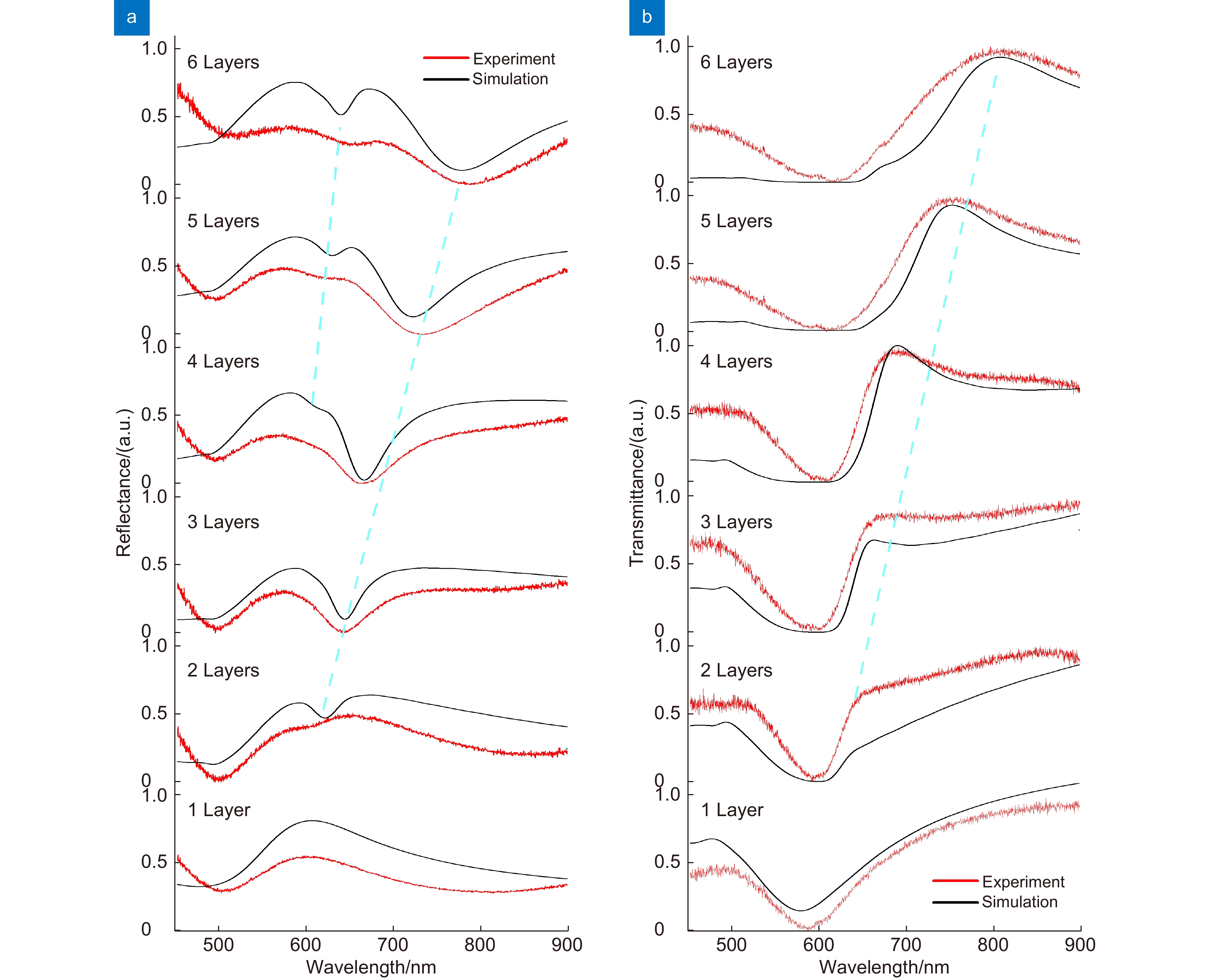
图 2 尺寸为20 nm的金纳米颗粒薄膜,从一层到六层的的反射光谱(a)和透射光谱(b),黑色实线表示仿真模拟结果,红色线条表示实验测量结果
Figure 2. Reflectance spectra (a) and transmission spectra (b) of gold nanoparticle films with a size of 20 nm, from one to six layers, with the solid black line indicating the simulation results and the red line indicating the experimental measurements
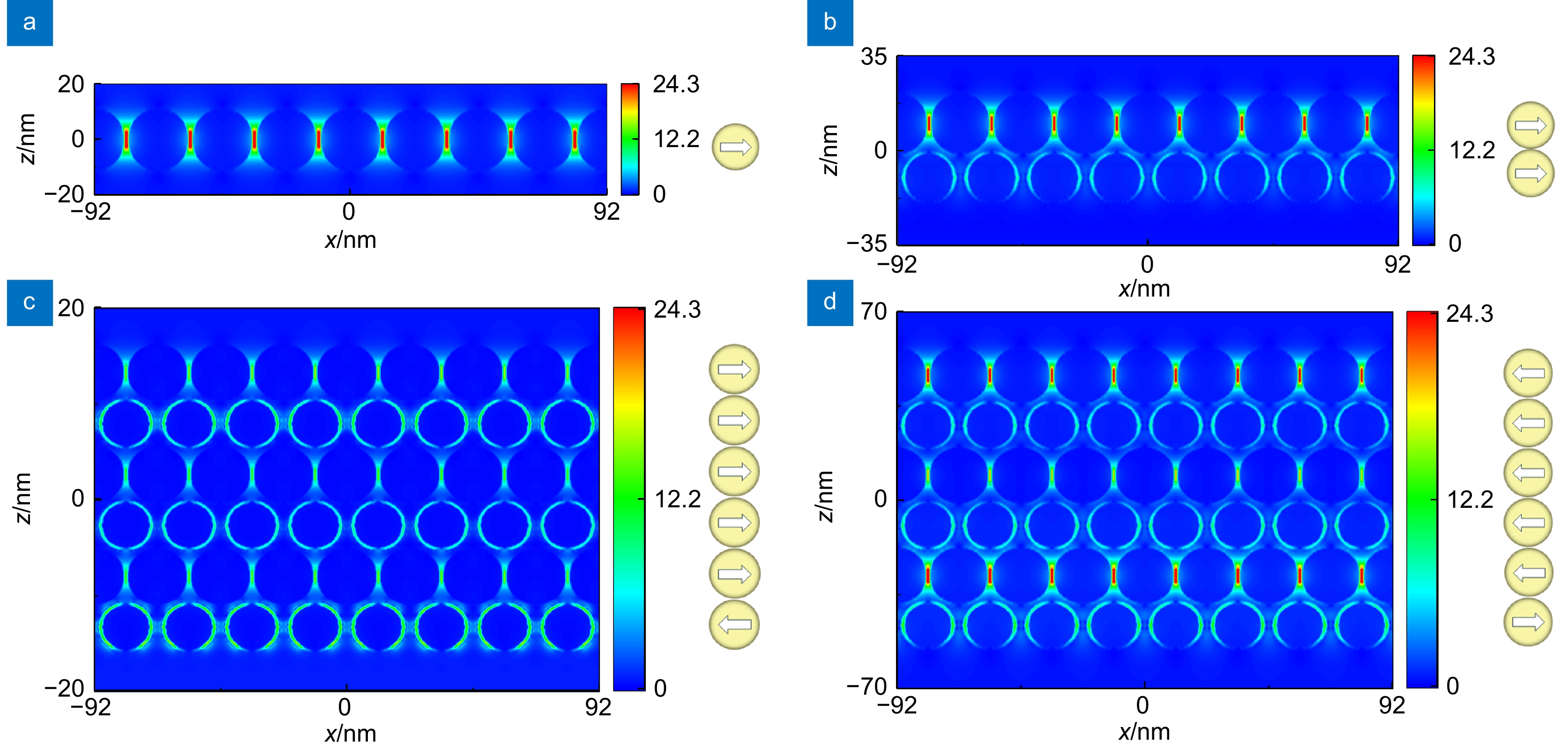
图 3 直径为20 nm,颗粒间间隔为1 nm的金纳米颗粒薄膜近场分布图:(a)单层;(b)双层;(c)六层(λ=638 nm);(d)六层(λ=778 nm)。在旁边展示了通过纳米颗粒内部的电流分布分配等离子体模式,每层颗粒中的偶极矩由白色箭头指示
Figure 3. Near-field distribution of a thin film of gold nanoparticles with a diameter of 20 nm and an interparticle spacing of 1 nm in (a) a single layer, (b) a bilayer, (c) a six-layer (λ = 638 nm), and (d) a six-layer (λ = 778 nm). The plasma pattern of current distribution through the interior of the nanoparticles is demonstrated next to it, and the dipole moments in the particles of each layer are indicated by the white arrows
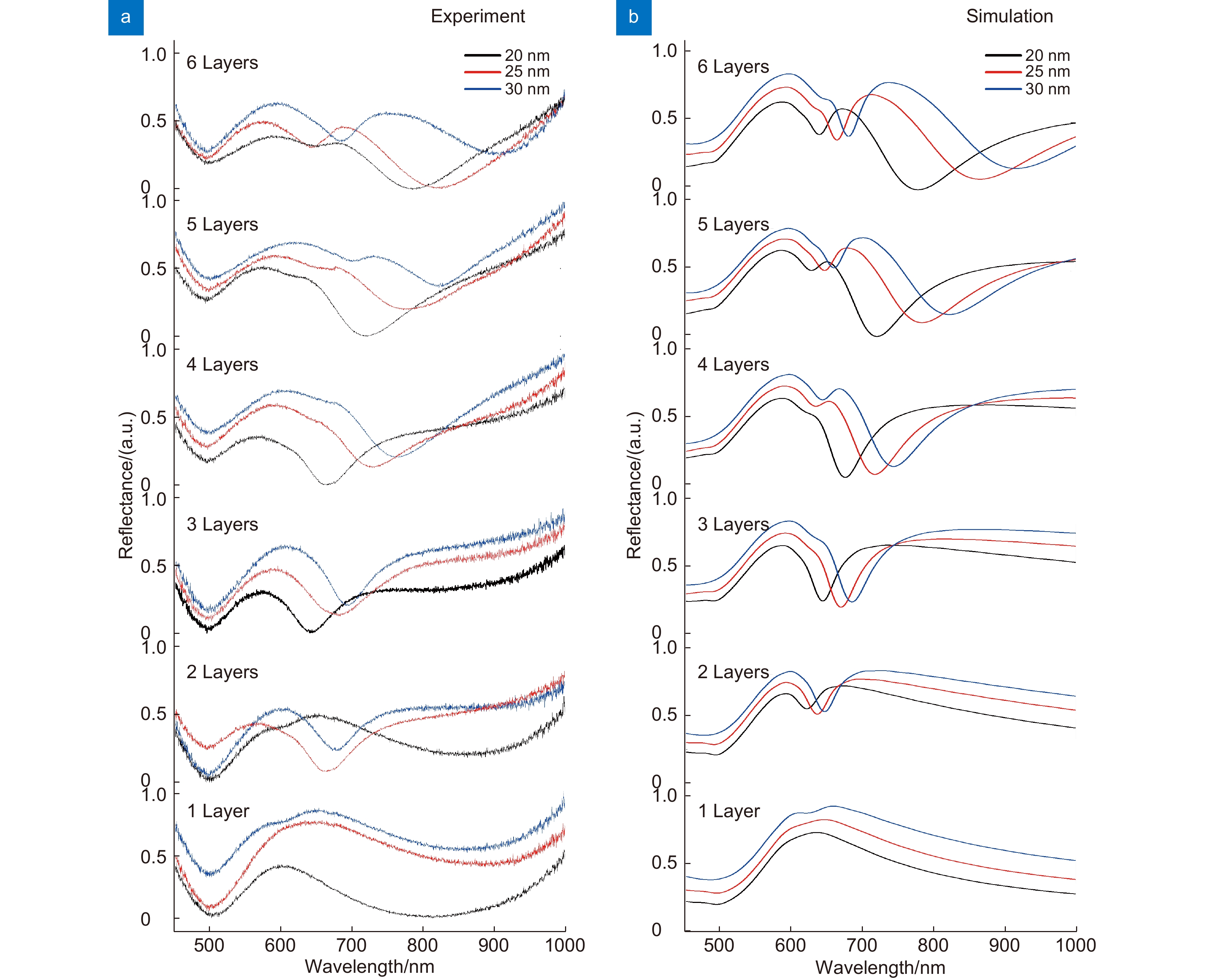
图 4 不同直径的(20 nm (黑色曲线)、25 nm (红色曲线)、30 nm (蓝色曲线))金纳米颗粒超晶格薄膜不同层数的反射光谱。 (a)实验测量结果; (b)数值计算结果
Figure 4. Reflectance spectra of gold nanoparticle superlattice films with different numbers of layers and diameters (20 nm - black curve, 25 nm - red curve, 30 nm - blue curve). (a) Experimental measurement results; (b) Numerical simulation results
表 1 不同尺寸金纳米颗粒的溶剂使用量
Table 1. Solvent usage of gold nanoparticles of different sizes
金球尺寸
/nm金球浓度
/(nmol/L)金球体积
/mL混合溶液
/mLPFT
/(mg/mL)18 4 2.25 4.5 10 20 3.3 2.25 4.5 10 22 2.6 2.25 4.5 10 25 1.8 2.25 4.5 10 30 1.4 2.25 4.5 10  下载: 导出CSV
-
[1] García-Lojo D, Núñez-Sánchez S, Gómez-Graña S, et al. Plasmonic supercrystals[J]. Acc Chem Res, 2019, 52(7): 1855−1864. doi: 10.1021/acs.accounts.9b00213
[2] Ross M B, Mirkin C A, Schatz G C. Optical properties of one-, two-, and three-dimensional arrays of plasmonic nanostructures[J]. J. Phys Chem C, 2016, 120(2): 816−830. doi: 10.1021/acs.jpcc.5b10800
[3] Mueller N S, Okamura Y, Vieira B G M, et al. Deep strong light–matter coupling in plasmonic nanoparticle crystals[J]. Nature, 2020, 583(7818): 780−784. doi: 10.1038/s41586-020-2508-1
[4] Solís D M, Taboada J M, Obelleiro F, et al. Toward ultimate nanoplasmonics modeling[J]. ACS Nano, 2014, 8(8): 7559−7570. doi: 10.1021/nn5037703
[5] Blanco-Formoso M, Pazos-Perez N, Alvarez-Puebla R A. Fabrication of plasmonic supercrystals and their SERS enhancing properties[J]. ACS Omega, 2020, 5(40): 25485−25492. doi: 10.1021/acsomega.0c03412
[6] Palmer S J, Xiao X F, Pazos-Perez N, et al. Extraordinarily transparent compact metallic metamaterials[J]. Nat Commun, 2019, 10(1): 2118. doi: 10.1038/s41467-019-09939-8
[7] Sun S H, Murray C B, Weller D, et al. Monodisperse FePt nanoparticles and ferromagnetic FePt nanocrystal superlattices[J]. Science, 2000, 287(5460): 1989−1992. doi: 10.1126/science.287.5460.1989
[8] Shevchenko E V, Ringler M, Schwemer A, et al. Self-assembled binary superlattices of CdSe and Au nanocrystals and their fluorescence properties[J]. J Am Chem Soc, 2008, 130(11): 3274−3275. doi: 10.1021/ja710619s
[9] Bigioni T P, Lin X M, Nguyen T T, et al. Kinetically driven self assembly of highly ordered nanoparticle monolayers[J]. Nature Materials, 2006, 5(4): 265−270. doi: 10.1038/nmat1611
[10] Vieira B G M, Mueller N S, Barros E B, et al. Plasmonic properties of close-packed metallic nanoparticle mono- and bilayers[J]. J Phys Chem C, 2019, 123(29): 17951−17960. doi: 10.1021/acs.jpcc.9b03859
[11] Mueller N S, Vieira B G M, Höing D, et al. Direct optical excitation of dark plasmons for hot electron generation[J]. Faraday Discuss, 2019, 214(214): 159−173. doi: 10.1039/C8FD00149A
[12] Mueller N S, Vieira B G M, Schulz F, et al. Dark interlayer plasmons in colloidal gold nanoparticle bi- and few-layers[J]. ACS Photonics, 2018, 5(10): 3962−3969. doi: 10.1021/acsphotonics.8b00898
[13] Tong J C, Suo F, Ma J H Z, et al. Surface plasmon enhanced infrared photodetection[J]. Opto-Electron Adv, 2019, 2(1): 180026. doi: 10.29026/oea.2019.180026
[14] Chiu C Y, Chen C K, Chang C W, et al. Surfactant-directed fabrication of supercrystals from the assembly of polyhedral Au-Pd core-shell nanocrystals and their electrical and optical properties[J]. J Am Chem Soc, 2015, 137(6): 2265−2275. doi: 10.1021/ja509044q
[15] Gómez-Graña S, Le Beulze A. Hierarchical self-assembly of a bulk metamaterial enables isotropic magnetic permeability at optical frequencies[J]. Mater Horiz, 2016, 3(6): 596−601. doi: 10.1039/C6MH00270F
[16] Alvarez-Puebla R A, Agarwal A, Manna P, et al. Gold nanorods 3D-supercrystals as surface enhanced Raman scattering spectroscopy substrates for the rapid detection of scrambled prions[J]. Proceedings of the National Academy of Sciences of the United States of America, 2011, 108(20): 8157−8161. doi: 10.1073/pnas.1016530108
[17] Alba M, Pazos-Perez N, Vaz B, et al. Macroscale plasmonic substrates for highly sensitive surface-enhanced Raman scattering[J]. Angew Chem Int Ed, 2013, 52(25): 6459−6463. doi: 10.1002/anie.201302285
[18] Yi C L, Liu H, Zhang S Y, et al. Self-limiting directional nanoparticle bonding governed by reaction stoichiometry[J]. Science, 2020, 369(6509): 1369−1374. doi: 10.1126/science.aba8653
[19] Chakraborty I N, Roy P, Rao A, et al. The unconventional role of surface ligands in dictating the light harvesting properties of quantum dots[J]. J Mater Chem A, 2021, 9(12): 7422−7457. doi: 10.1039/D0TA12623C
[20] Roy P, Devatha G, Roy S, et al. Electrostatically driven resonance energy transfer in an all-quantum dot based donor-acceptor system[J]. J Phys Chem Lett, 2020, 11(13): 5354−5360. doi: 10.1021/acs.jpclett.0c01360
[21] Bai P, Yang S, Bao W, et al. Diversifying nanoparticle assemblies in supramolecule nanocomposites via cylindrical confinement[J]. Nano Lett, 2017, 17(11): 6847−6854. doi: 10.1021/acs.nanolett.7b03131
[22] Macfarlane R J, Lee B, Jones M R, et al. Nanoparticle superlattice engineering with DNA[J]. Science, 2011, 334(6053): 204−208. doi: 10.1126/science.1210493
[23] Auyeung E, Li T I N G, Senesi A J, et al. DNA-mediated nanoparticle crystallization into Wulff polyhedra[J]. Nature, 2014, 505(7481): 73−77. doi: 10.1038/nature12739
[24] Schulz F, Pavelka O, Lehmkühler F, et al. Structural order in plasmonic superlattices[J]. Nat Commun, 2020, 11(1): 3821. doi: 10.1038/s41467-020-17632-4
[25] Song L P, Xu B B, Cheng Q, et al. Instant interfacial self-assembly for homogeneous nanoparticle monolayer enabled conformal “lift-on” thin film technology[J]. Sci Adv, 2021, 7(52): eabk2852. doi: 10.1126/sciadv.abk2852
[26] Zheng Y Q, Zhong X L, Li Z Y, et al. Successive, seed-mediated growth for the synthesis of single-crystal gold nanospheres with uniform diameters controlled in the range of 5–150 nm[J]. Part Part Syst Char, 2014, 31(2): 266−273. doi: 10.1002/ppsc.201300256
[27] Sun Q, Aguila B, Perman J A, et al. Integrating superwettability within covalent organic frameworks for functional coating[J]. Chem, 2018, 4(7): 1726−1739. doi: 10.1016/j.chempr.2018.05.020
[28] 韩莹莹, 陈盼盼, 王曼, 等. 基于悬链线纳米粒子超构表面的线偏振光SPPs定向激发[J]. 光电工程, 2022, 49(10): 220105. doi: 10.12086/oee.2022.220105
Han Y Y, Chen P P, Wang M, et al. SPPs directional excitation of linearly polarized light based on catenary nanoparticle metasurface[J]. Opto-Electron Eng, 2022, 49(10): 220105. doi: 10.12086/oee.2022.220105
-
 点击扫一扫
点击扫一扫
图(5)
表(1)
计量
- 文章访问数:
- PDF下载数:
- 施引文献: 0


