
 E-mail Alert
 RSS
RSS
Image quality optimization of line-focused spectral domain optical coherence tomography with subsection dispersion compensation
-
摘要:
本研究中搭建了一套用于生物样品成像的线聚焦谱域光学相干层析(LF-SD-OCT)系统,针对系统误差引起的轴向展宽和灵敏度衰减问题,提出了一种分段色散补偿的方法,对成像深度内的二阶和三阶色散相位进行了补偿,并通过平面镜和透明胶带样品成像实验来验证该方法的有效性和可靠性。通过本文提出的分段色散补偿方法和其他成熟方法的联合应用,系统在获得57.2 kHz的等效A-scan速率的同时,轴向分辨率提升至6.76 μm,对胶带样品2 mm深度范围内及水果样品0.3 mm深度范围内进行了较清晰成像。实验结果表明,该方法可以在兼顾处理效率的同时提高全深度轴向分辨率和灵敏度,未来有望广泛实现线聚焦谱域光学相干层析的生物医学成像应用。
Abstract:In this study, a line-focused spectral domain optical coherence tomography (LF-SD-OCT) system for imaging biological samples was built, and a data processing algorithm to improve the imaging quality was proposed to solve most of the problems of axial broadening and sensitivity attenuation caused by systematic errors. In particular, a segmented dispersion compensation method is proposed to compensate the second- and third-order dispersion phases in the imaging depth. The effectiveness and reliability of this method are verified by the imaging experiments of plane mirrors and scotch tape samples. Finally, it is proved that this method can improve the full-depth axial resolution and sensitivity without affecting the image processing speed. The final system can achieve the axial resolution of 6.76 μm and an equivalent A-scan rate of 57.2 kHz, and clearly image the tape sample within 2 mm depth and the apple sample within 0.3 mm depth. In the future, it is expected to widely realize the biological imaging applications of line-focused spectral domain optical coherence tomography.
-
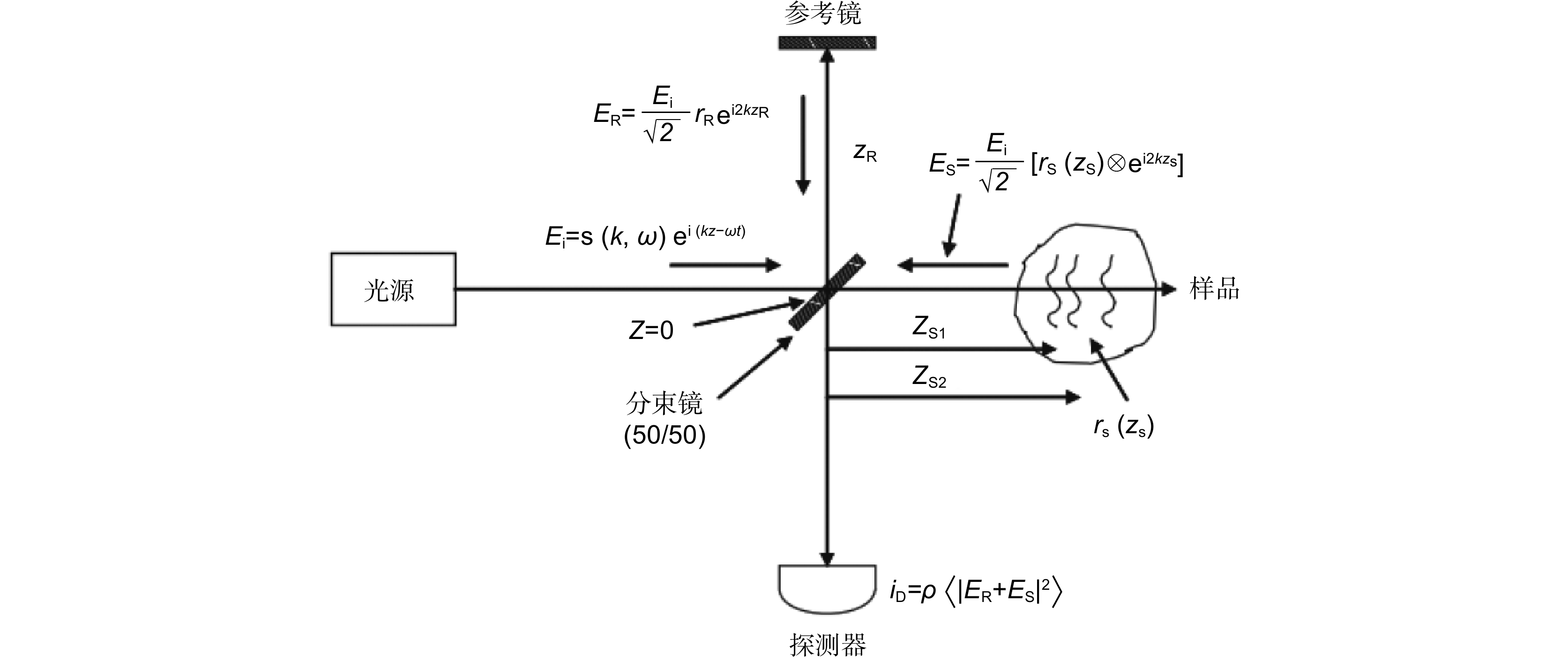
Overview: LF-SD-OCT was first proposed by Zuluaga and Kortum et al in 1999 and has since made significant advances in imaging speed and resolution. Compared with mainstream SD-OCT, LF-SD-OCT has advantages in system cost, imaging speed, and biosecurity, so it has a very good application prospect in areas with high imaging speed and security requirements, such as in vivo imaging. However, as the depth of LF-SD-OCT increases, the image quality deteriorates rapidly. At present, LF-SD-OCT has not been successfully applied in the biomedical field in China, and there are only a few successful cases in the international community.
In this study, we independently designed and built a line-focused spectral domain optical coherence tomography (LF-SD-OCT) system for imaging biological samples, and proposed data processing algorithms to improve imaging quality. We found that the dispersion parameters of different depth positions are quite different, and the unified dispersion compensation coefficient will lead to undercompensation or overcompensation in some regions. First, the curve of the dispersion compensation coefficient with depth is obtained by system calibration. Then, the original data is divided into four segments in the depth direction, and the dispersion compensation coefficient corresponding to the center position of each segment is used to compensate the second- and third-order dispersion phase in the segment. Subsequently, the segments are combined. The effectiveness and reliability of the proposed method are verified by using a flat mirror and scotch tape sample imaging. Finally, it is proved that the proposed method can improve the full-depth axial resolution and sensitivity while taking into account the image processing speed. After iterative wavelength distribution calibration, piecewise dispersion compensation, and deconvolution denoising, most of the axial broadening and sensitivity attenuation problems caused by systematic errors have been solved. The final system can achieve the axial resolution of 6.76 μm (theoretical value is 6.2 μm) and an equivalent A-scan rate of 57.2 kHz, and can image the tape sample within 2 mm depth and the apple sample within 0.3 mm depth.
This study has proved that line-focused spectral OCT has great advantages in imaging speed and development cost. In the future, further increase of light source power and appropriate improvement of the optical path can image human samples with weaker backscattered light, and it is expected that line-focused spectral OCT can be successfully applied to domestic biomedical imaging fields.
-

-
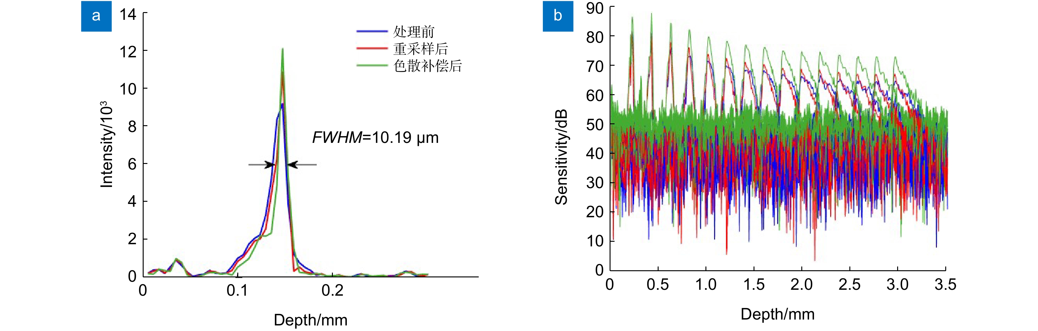
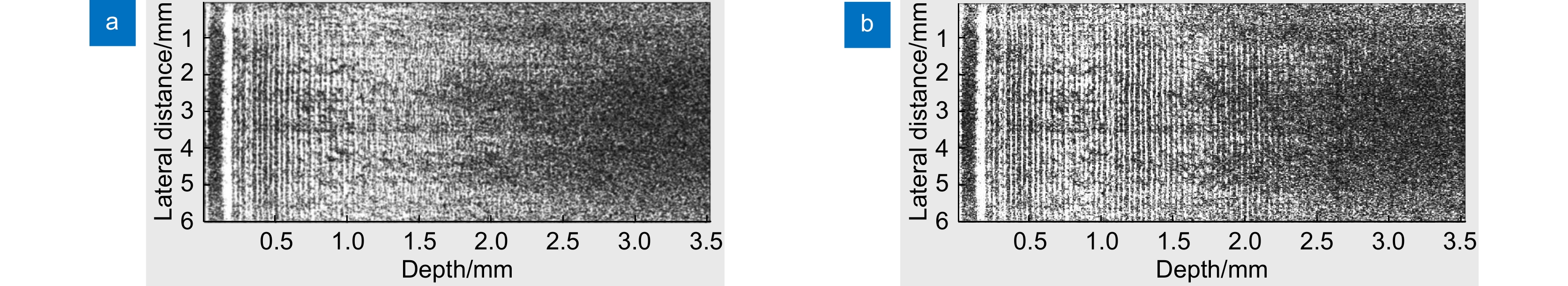
图 4 重采样和色散补偿前后对比图。 (a)轴向分辨率对比图; (b)系统滚降对比图
Figure 4. Comparison before and after resampling and dispersion compensation. (a) Comparison of axial resolution; (b) Comparison of system roll down
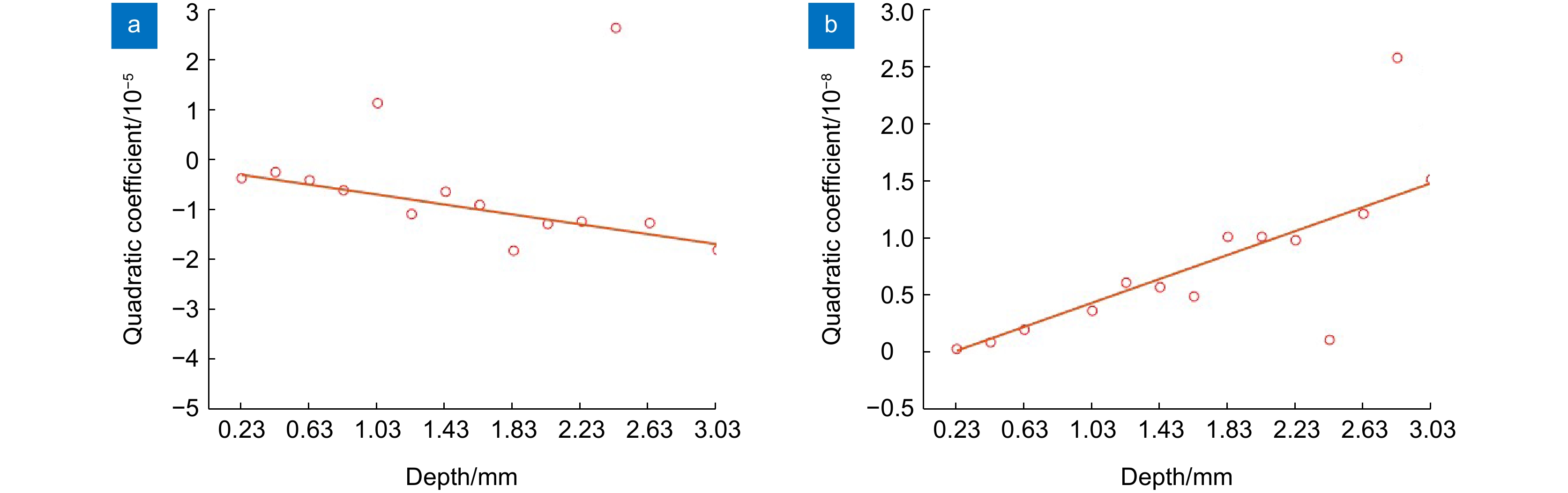
图 6 色散补偿系数随深度变化图。 (a) 二次项系数; (b) 三次项系数
Figure 6. Diagram of dispersion compensation coefficient with depth. (a) Quadratic coefficient; (b) Cubic coefficient
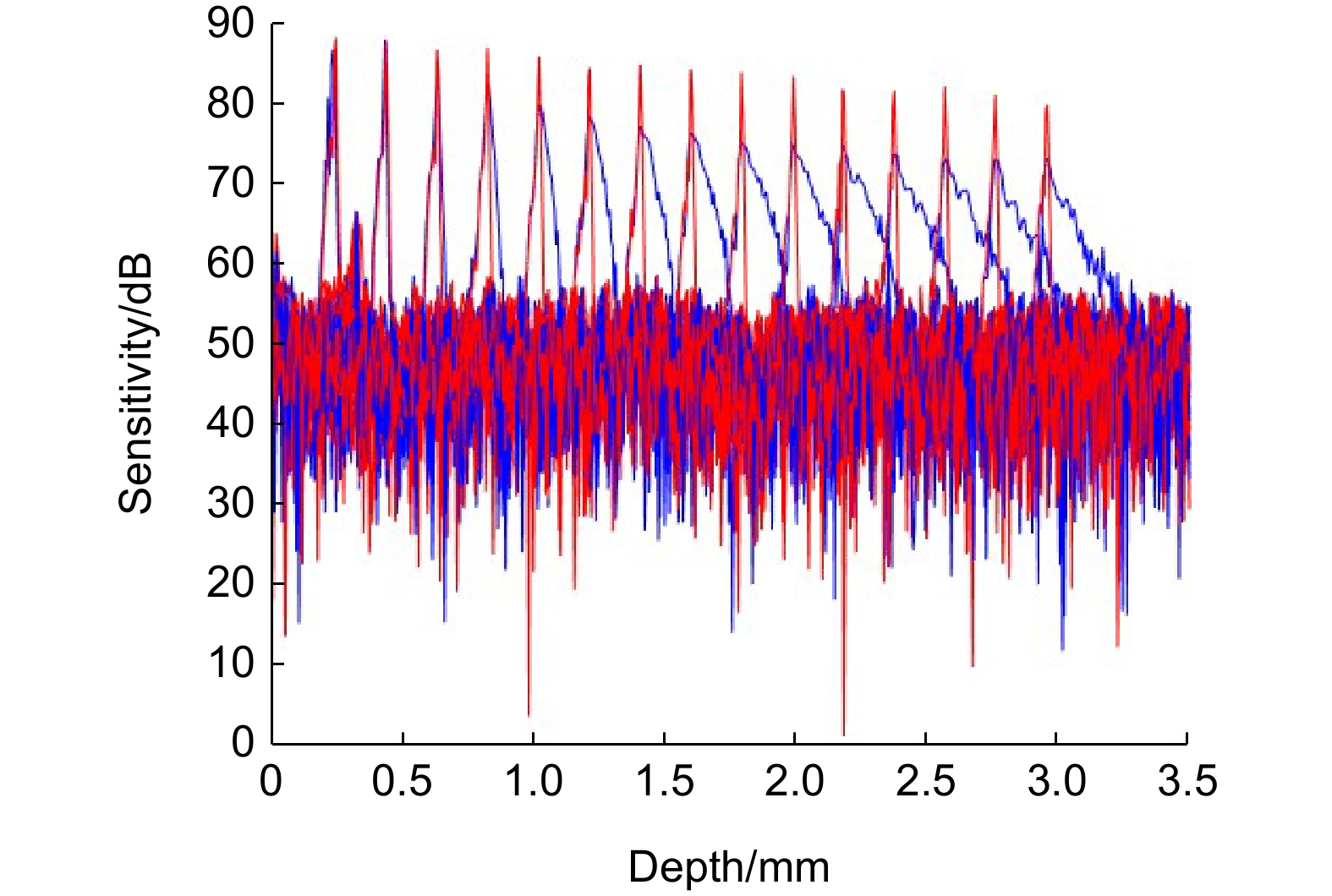
图 7 针对性色散补偿前 (蓝)后 (红)系统滚降对比图
Figure 7. Comparison of system roll-down before (blue) and after (red) targeted dispersion compensation
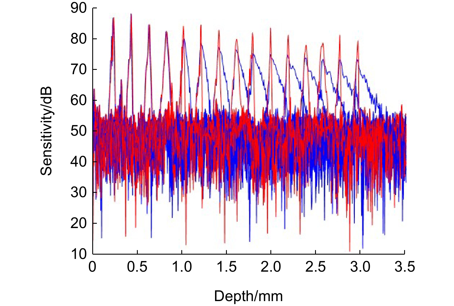
图 8 分段色散补偿前 (蓝)后 (红)系统滚降对比图
Figure 8. Comparison of system roll-down before (blue) and after (red) subsection dispersion compensation
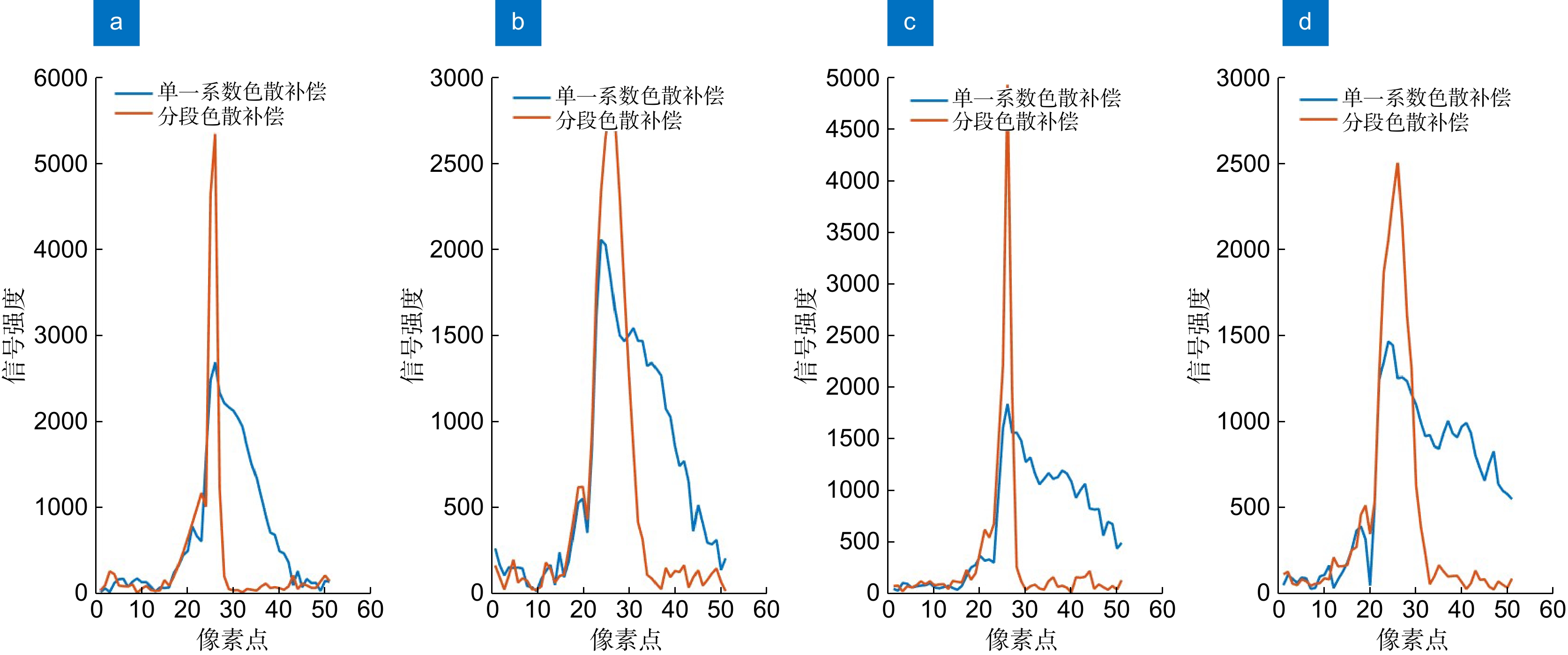
图 9 分段色散补偿前后成像结果对比图。 (a) Δz=1.25 mm; (b) Δz=1.65 mm; (c) Δz=2.05 mm; (d) Δz=2.45 mm
Figure 9. Comparison of imaging results before and after segmented dispersion compensation. (a) Δz=1.25 mm; (b) Δz=1.65 mm; (c) Δz=2.05 mm; (d) Δz=2.45 mm
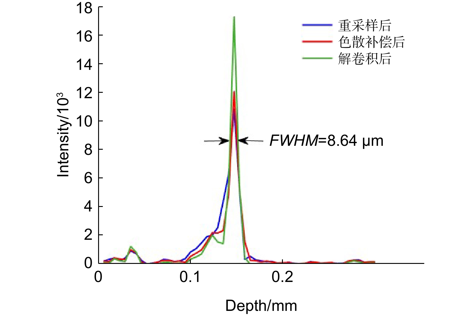
图 10 解卷积前 (蓝红)后 (绿)结果对比
Figure 10. Comparison of results before and after deconvolution
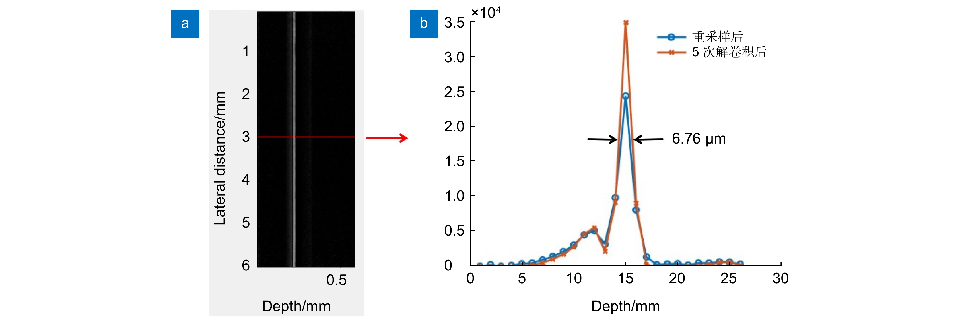
图 11 光程差约0.375 mm平面镜样品成像结果。 (a) 平面镜样品成像结果; (b) 线照明中心位置轴向分辨率
Figure 11. Flat mirror sample imaging results (Δz=0.375 mm). (a) Flat mirror sample imaging results; (b) Axial resolution of focus line center position
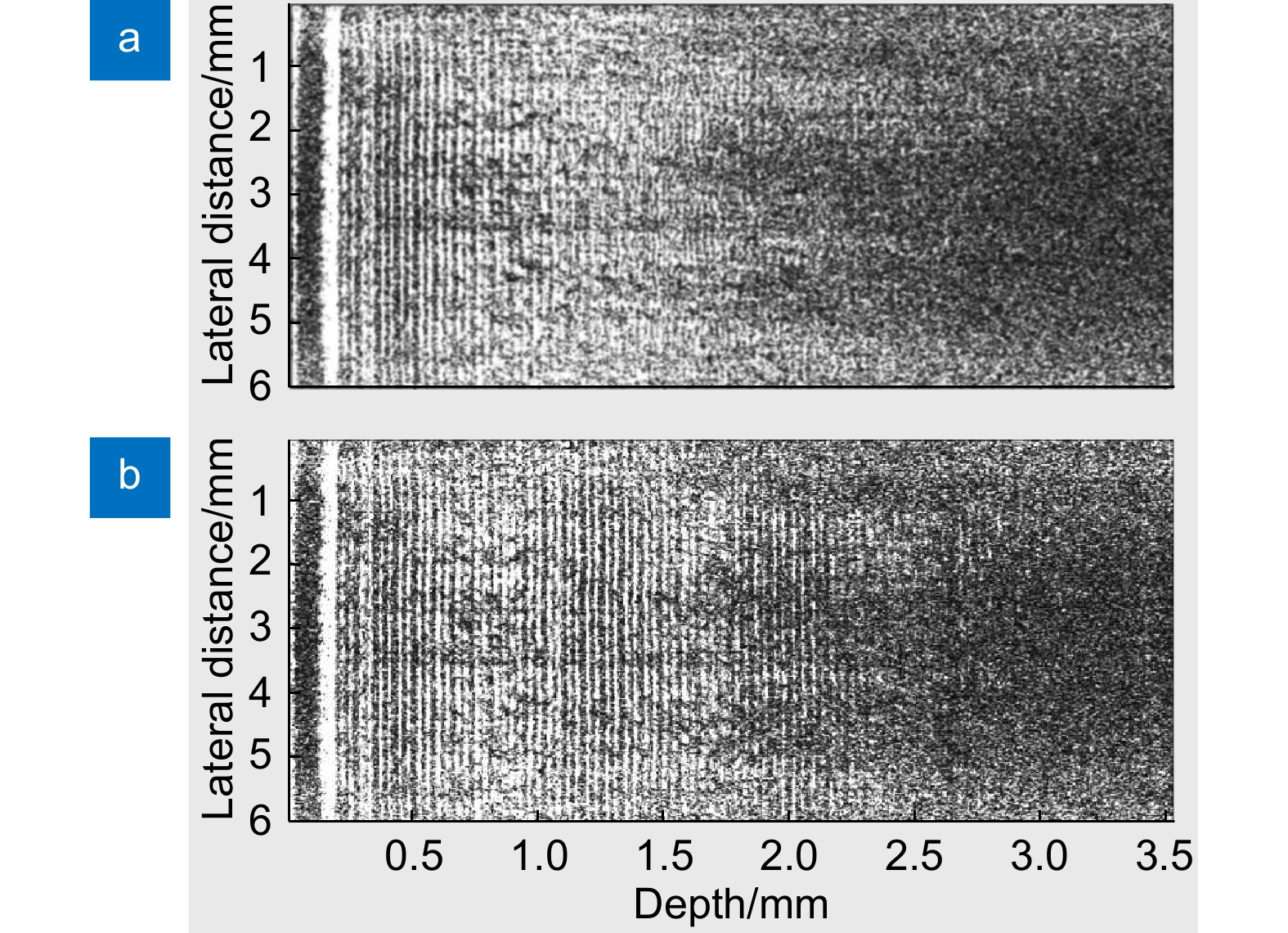
图 12 透明胶带成像结果。 (a) 单一系数色散补偿后图像; (b) 分段色散补偿后图像
Figure 12. Scotch tape imaging results. (a) Image after single coefficient dispersion compensation; (b) Image after subsection dispersion compensation
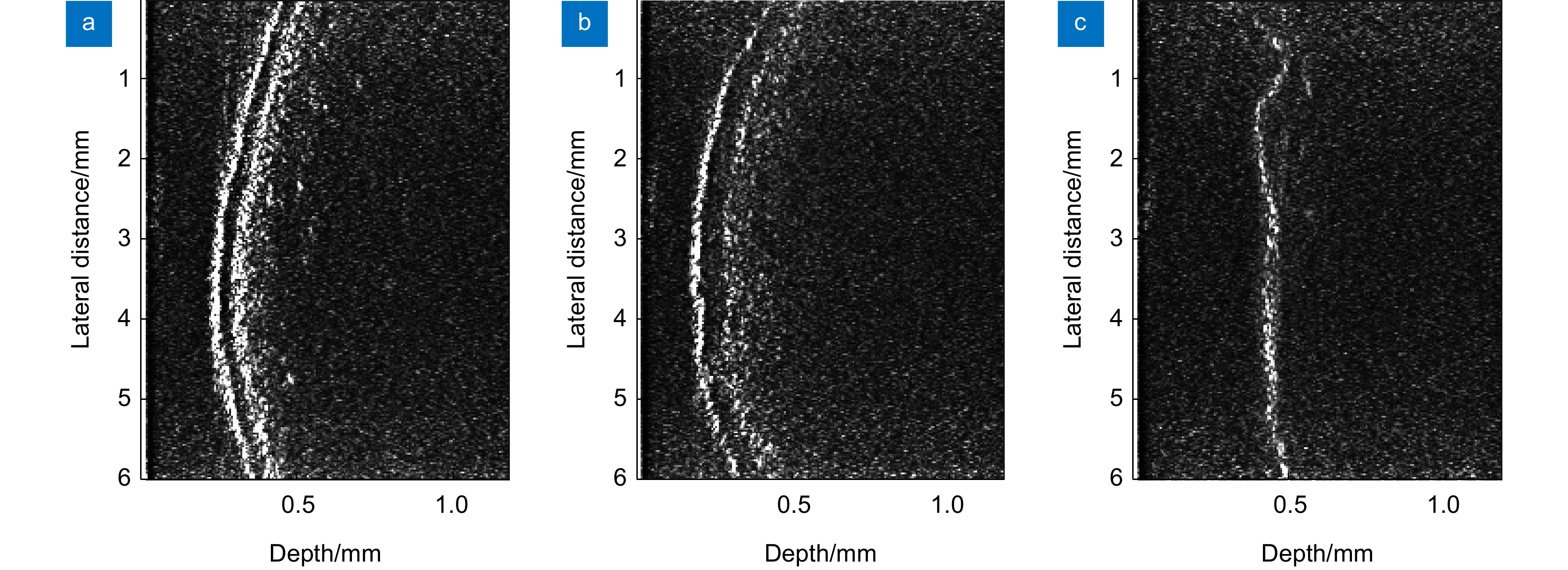
图 14 水果样品成像结果。 (a) 苹果表皮; (b) 梨表皮; (c) 橙子囊瓣
Figure 14. Fruits sample imaging results. (a) Apple skin; (b) Pear skin; (c) Orange saccule
表 1 大深度位置灵敏度和轴向分辨对比
Table 1. Comparison of sensitivity and axial resolution
OPD Δz/mm Axial resolution/μm Sensitivity/dB 1.05 44.95→12.9 79.4→84.6 1.25 56.60→10.98 78.6→84.7 1.45 68.50→17.40 77.1→82.7 1.65 84.05→36.05 76.3→79.0 1.85 89.10→12.15 75.8→82.6 2.05 98.40→8.67 75.3→83.9 2.25 97.66→18.53 74.5→80.5 2.45 125.89→35.41 73.3→78.0 2.65 123.45→37.15 72.8→76.3 2.85 130.18→11.25 72.4→81.7 3.05 96.11→21.12 73.1→79.3  下载: 导出CSV
-
[1] Huang D, Swanson E A, Lin C P, et al. Optical coherence tomography[J]. Science, 1991, 254(5035): 1178−1181. doi: 10.1126/science.1957169
[2] Fujimoto J G. Optical coherence tomography for ultrahigh resolution in vivo imaging[J]. Nat Biotechnol, 2003, 21(11): 1361−1367. doi: 10.1038/nbt892
[3] 刘逸飞, 苏亚, 姚晓天, 等. OCT无创血糖检测图像处理最优化方法研究[J]. 激光技术, 2023, 47(2): 178−184. doi: 10.7510/jgjs.issn.1001-3806.2023.02.004
Liu Y F, Su Y, Yao X T, et al. An optimization method of image processing for OCT non-invasive blood glucose detection[J]. Laser Technol, 2023, 47(2): 178−184. doi: 10.7510/jgjs.issn.1001-3806.2023.02.004
[4] Farazdaghi M K, Ebrahimi K B. Role of the choroid in age-related macular degeneration: a current review[J]. J Ophthalmic Vis Res, 2019, 14(1): 78−87. doi: 10.4103/jovr.jovr_125_18
[5] Choi W, Moult E M, Waheed N K, et al. Ultrahigh-speed, swept-source optical coherence tomography angiography in nonexudative age-related macular degeneration with geographic atrophy[J]. Ophthalmology, 2015, 122(12): 2532−2544. doi: 10.1016/j.ophtha.2015.08.029
[6] Potsaid B, Baumann B, Huang D, et al. Ultrahigh speed 1050nm swept source / Fourier domain OCT retinal and anterior segment imaging at 100, 000 to 400, 000 axial scans per second[J]. Opt Express, 2010, 18(19): 20029−20048. doi: 10.1364/OE.18.020029
[7] 李云耀, 樊金宇, 蒋天亮, 等. 光学相干层析技术在眼科手术导航方面的研究进展[J]. 光电工程, 2023, 50(1): 220027. doi: 10.12086/oee.2023.220027
Li Y Y, Fan J Y, Jiang T L, et al. Review of the development of optical coherence tomography imaging navigation technology in ophthalmic surgery[J]. Opto-Electron Eng, 2023, 50(1): 220027. doi: 10.12086/oee.2023.220027
[8] Fercher A F, Hitzenberger C K, Kamp G, et al. Measurement of intraocular distances by backscattering spectral interferometry[J]. Opt Commun, 1995, 117(1-2): 43−48. doi: 10.1016/0030-4018(95)00119-S
[9] de Boer J F, Cense B, Park B H, et al. Improved signal-to-noise ratio in spectral-domain compared with time-domain optical coherence tomography[J]. Opt Lett, 2003, 28(21): 2067−2069. doi: 10.1364/OL.28.002067
[10] Choma M A, Sarunic M V, Yang C, et al. Sensitivity advantage of swept source and Fourier domain optical coherence tomography[J]. Opt Express, 2003, 11(18): 2183−2189. doi: 10.1364/OE.11.002183
[11] An L, Li P, Shen T T, et al. High speed spectral domain optical coherence tomography for retinal imaging at 500, 000 A-lines per second[J]. Biomed Opt Express, 2011, 2(10): 2770−2783. doi: 10.1364/BOE.2.002770
[12] Kocaoglu O P, Turner T L, Liu Z L, et al. Adaptive optics optical coherence tomography at 1 MHz[J]. Biomed Opt Express, 2014, 5(12): 4186−4200. doi: 10.1364/BOE.5.004186
[13] Choi D H, Hiro-Oka H, Shimizu K, et al. Spectral domain optical coherence tomography of multi-MHz A-scan rates at 1310 nm range and real-time 4D-display up to 41 volumes/second[J]. Biomed Opt Express, 2012, 3(12): 3067−3086. doi: 10.1364/BOE.3.003067
[14] Wang R K, An L. Multifunctional imaging of human retina and choroid with 1050-nm spectral domain optical coherence tomography at 92-kHz line scan rate[J]. J Biomed Opt, 2011, 16(5): 050503. doi: 10.1117/1.3582159
[15] Seong D, Jeon D, Wijesinghe R E, et al. Ultrahigh-speed spectral-domain optical coherence tomography up to 1-MHz a-scan rate using space–time-division multiplexing[J]. IEEE Trans Instrum Meas, 2021, 70: 4504108. doi: 10.1109/TIM.2021.3073701
[16] Zuluaga A F, Richards-Kortum R. Spatially resolved spectral interferometry for determination of subsurface structure[J]. Opt Lett, 1999, 24(8): 519−521. doi: 10.1364/OL.24.000519
[17] Han L, Hosseiaee Z, Tan B, et al. High resolution line-field SD-OCT with 2.5 kHz frame rate for cellular resolution imaging of biological tissue[J]. Proc SPIE, 2019, 10867: 108672X. doi: 10.1117/12.2511686
[18] Lawman S, Mason S, Kaye S B, et al. Accurate in vivo bowman's thickness measurement using mirau ultrahigh axial resolution line field optical coherence tomography[J]. Transl Vis Sci Technol, 2022, 11(8): 6. doi: 10.1167/TVST.11.8.6
[19] 沈毅, 陈志彦, 邱建榕, 等. 并行谱域光学相干层析成像技术的研究进展[J]. 中国激光, 2018, 45(2): 0207004. doi: 10.3788/CJL201845.0207004
Shen Y, Chen Z Y, Qiu J R, et al. Research progress on parallel spectral domain optical coherence tomography technology[J]. Chin J Lasers, 2018, 45(2): 0207004. doi: 10.3788/CJL201845.0207004
[20] Nakamura Y, Makita S, Yamanari M, et al. High-speed three-dimensional human retinal imaging by line-field spectral domain optical coherence tomography[J]. Opt Express, 2007, 15(12): 7103−7116. doi: 10.1364/OE.15.007103
[21] Grajciar B, Lehareinger Y, Fercher A F, et al. High sensitivity phase mapping with parallel Fourier domain optical coherence tomography at 512 000 A-scan/s[J]. Opt Express, 2010, 18(21): 21841−21850. doi: 10.1364/OE.18.021841
[22] Drexler W, Fujimoto J G. Optical Coherence Tomography: Technology and Applications[M]. New York: Springer, 2008.
[23] Hitzenberger C K, Baumgartner A, Drexler W, et al. Dispersion effects in partial coherence interferometry: implications for intraocular ranging[J]. J Biomed Opt, 1999, 4(1): 144−151. doi: 10.1117/1.429900
[24] 黄炳杰, 步鹏, 王向朝, 等. 用于频域光学相干层析成像的深度分辨色散补偿方法[J]. 光学学报, 2012, 32(2): 0217002. doi: 10.3788/AOS201232.0217002
Huang B J, Bu P, Wang X Z, et al. Optical coherence tomography based on depth resolved dispersion compensation[J]. Acta Opt Sin, 2012, 32(2): 0217002. doi: 10.3788/AOS201232.0217002
[25] Marks D L, Oldenburg A L, Reynolds J J, et al. Autofocus algorithm for dispersion correction in optical coherence tomography[J]. Appl Opt, 2003, 42(16): 3038−3046. doi: 10.1364/AO.42.003038
[26] Wojtkowski M, Srinivasan V J, Ko T H, et al. Ultrahigh-resolution, high-speed, Fourier domain optical coherence tomography and methods for dispersion compensation[J]. Opt Express, 2004, 12(11): 2404−2422. doi: 10.1364/OPEX.12.002404
[27] Diddams S, Diels J C. Dispersion measurements with white-light interferometry[J]. J Opt Soc Am B, 1996, 13(6): 1120−1129. doi: 10.1364/JOSAB.13.001120
[28] Wang K, Ding Z H. Spectral calibration in spectral domain optical coherence tomography[J]. Chin Opt Lett, 2008, 6(12): 902−904. doi: 10.3788/COL20080612.0902
[29] 潘柳华, 李中梁, 王向朝, 等. 光学相干层析成像随深度变化的色散补偿方法[J]. 光学学报, 2017, 37(5): 0511002. doi: 10.3788/AOS201737.0511002
Pan L H, Li Z L, Wang X Z, et al. Depth-dependent dispersion compensation for optical coherence tomography[J]. Acta Opt Sin, 2017, 37(5): 0511002. doi: 10.3788/AOS201737.0511002
[30] Liu Y H, Liang Y M, Mu G G, et al. Deconvolution methods for image deblurring in optical coherence tomography[J]. J Opt Soc Am A, 2009, 26(1): 72−77. doi: 10.1364/JOSAA.26.000072
-


 点击扫一扫
点击扫一扫
图(15)
表(1)
计量
- 文章访问数:
- PDF下载数:
- 施引文献: 0


