
 E-mail Alert
 RSS
RSS
-
摘要:
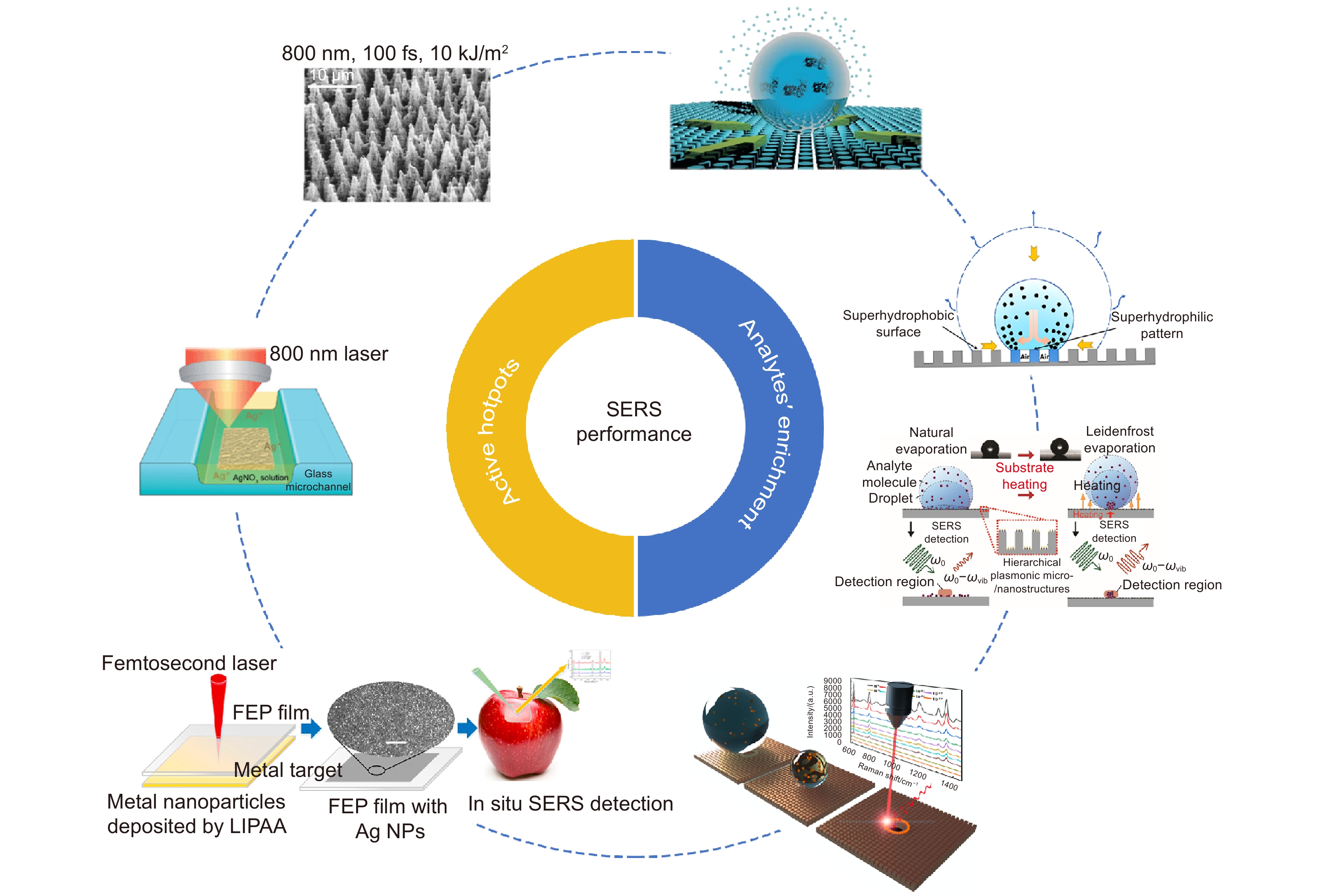
表面增强拉曼散射(SERS)传感器在诸多领域拥有重要的应用潜力。为实现高精度SERS检测,增加热点密度和热点区域中分析物分子数量成为当前研究的重点。超快激光可快速在材料表面构筑大面积的微纳米结构,对于高性能SERS基底的商业化制备具有重要的意义。本文从热点密度和检测区域中分析物分子浓度两个方面,总结了近年来超快激光制造高性能SERS基底的工艺方法。超快激光既能“自下而上”,也能“自上而下”加工出具有局域场增强效应的微纳米结构。其中,超快激光制备的超疏水表面是目前实现待测分子富集的有效方法之一。最后展望了激光制备SERS基底的应用前景。
Abstract:Surface-enhanced Raman scattering (SERS) provides important applications in diverse fields. In order to achieve high-precision SERS detection of trace molecules, current research focuses on how to increase the density of hot spots and the number of analyte molecules in the detection area. An ultrafast laser can rapidly construct large-area micro/nano-structures on material surfaces. It is important for the commercial preparation of high-performance SERS sensors. In this paper, the ultrafast laser preparation of high-performance SERS sensors is introduced from the aspect of the density of hot spots and the number of analyte molecules in the detection region. Ultrafast lasers enable both "bottom-up" and "top-down" processing. In particular, the superhydrophobic surface prepared by the ultrafast laser is one of the most effective methods to achieve the enrichment of analyte molecules. Finally, a prospect for the development of laser-prepared SERS substrates is provided.
-

-
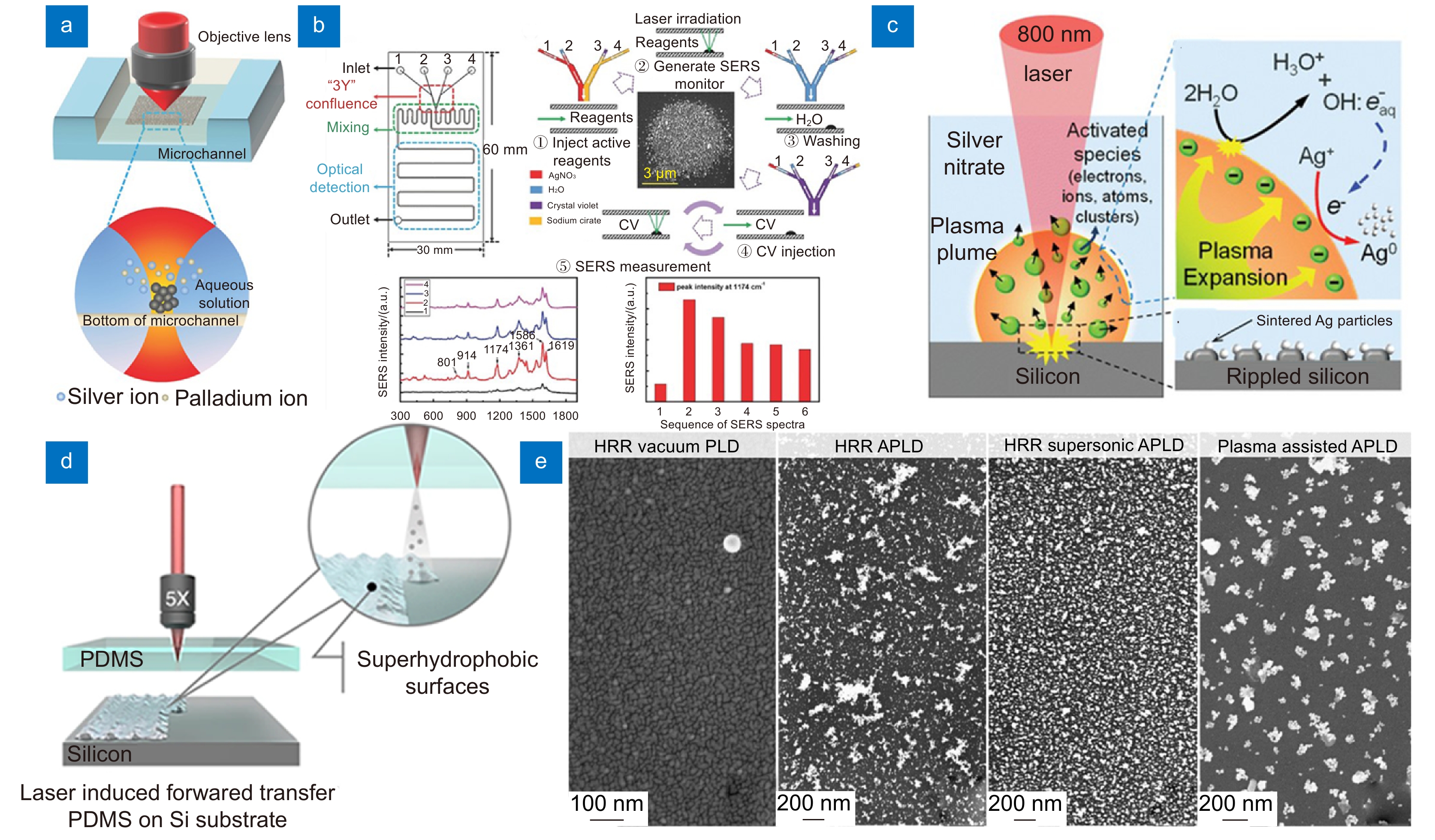
图 2 (a) 飞秒激光诱导银/钯合金还原示意图 [31]; (b) 两步飞秒激光诱导银离子还原形成的纳米结构 [32];(c) 固液界面等离子体增强还原银离子示意图 [33]; (d) 激光诱导正向转移制备SERS基片示意图 [35];(e) 脉冲激光沉积形成的金属纳米结构 [37]
Figure 2. (a) Schematic diagram of femtosecond laser-induced reduction of silver/palladium alloy [31]; (b) Nanostructures formed by two-step femtosecond laser-induced reduction of silver ions [32]; (c) Schematic diagram of the plasma-enhanced reduction of silver ionsat the solid-liquid interface [33]; (d) Schematic diagram of SERS substrates prepared by laser-induced forward transfer [35]; (e) Metal nanostructures formed by pulsed laser deposition [37]
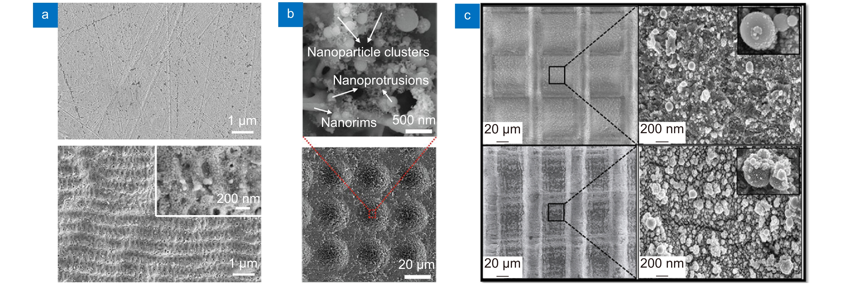
图 3 (a)飞秒激光在空气中制备银基SERS基底 [42]; (b) 飞秒激光在氩气氛围中制备银基SERS基底(S-Ag-Ar) [44];(c) 飞秒激光制备金基SERS基底 [47]
Figure 3. (a) Femtosecond laser preparation of silver-based SERS substrates in air [42]; (b) Femtosecond laser preparation of silver-based SERS substrates (S-Ag-Ar) in an argon atmosphere [44]; (c) Femtosecond laser preparation of gold-based SERS substrates [47]
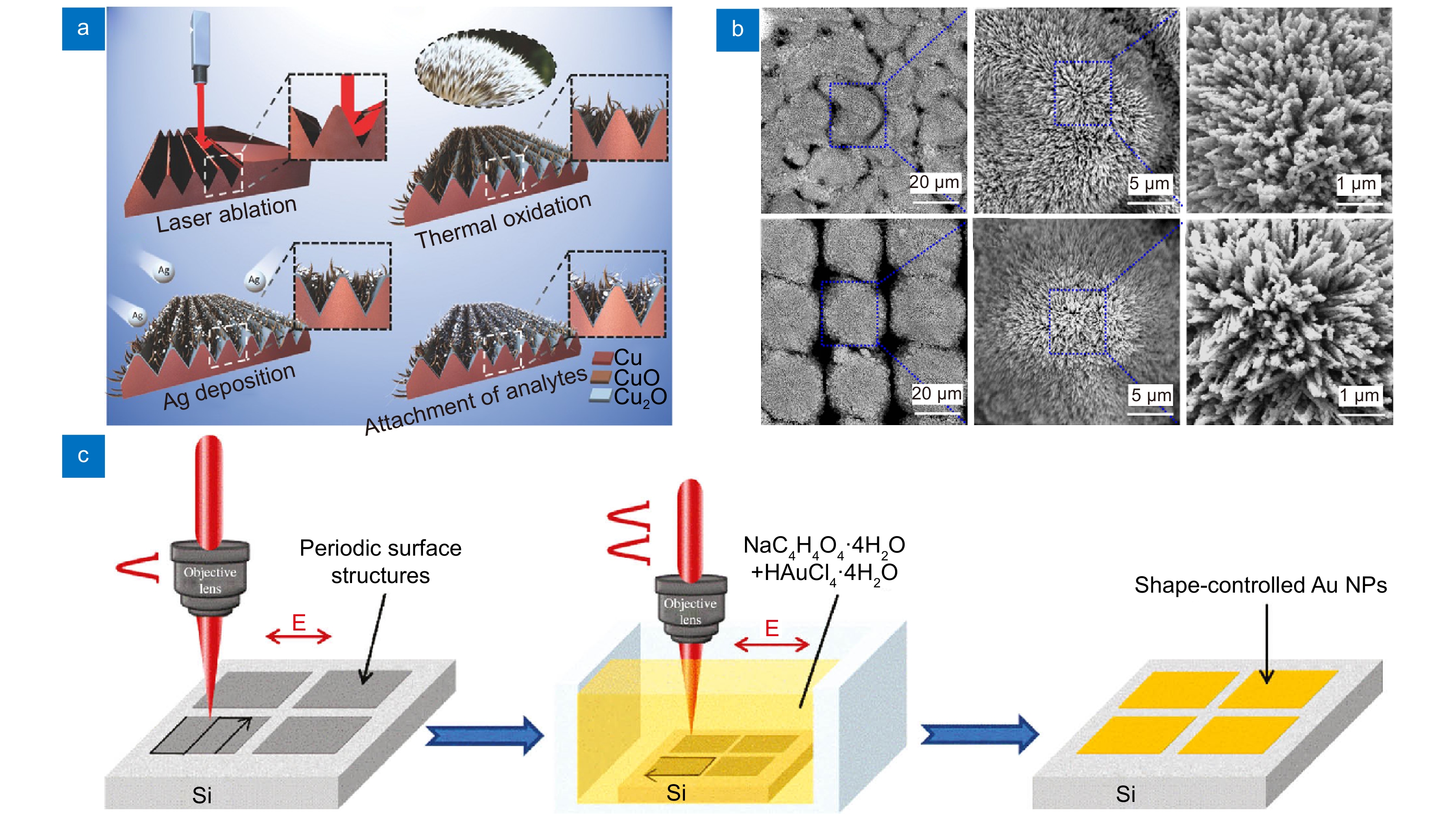


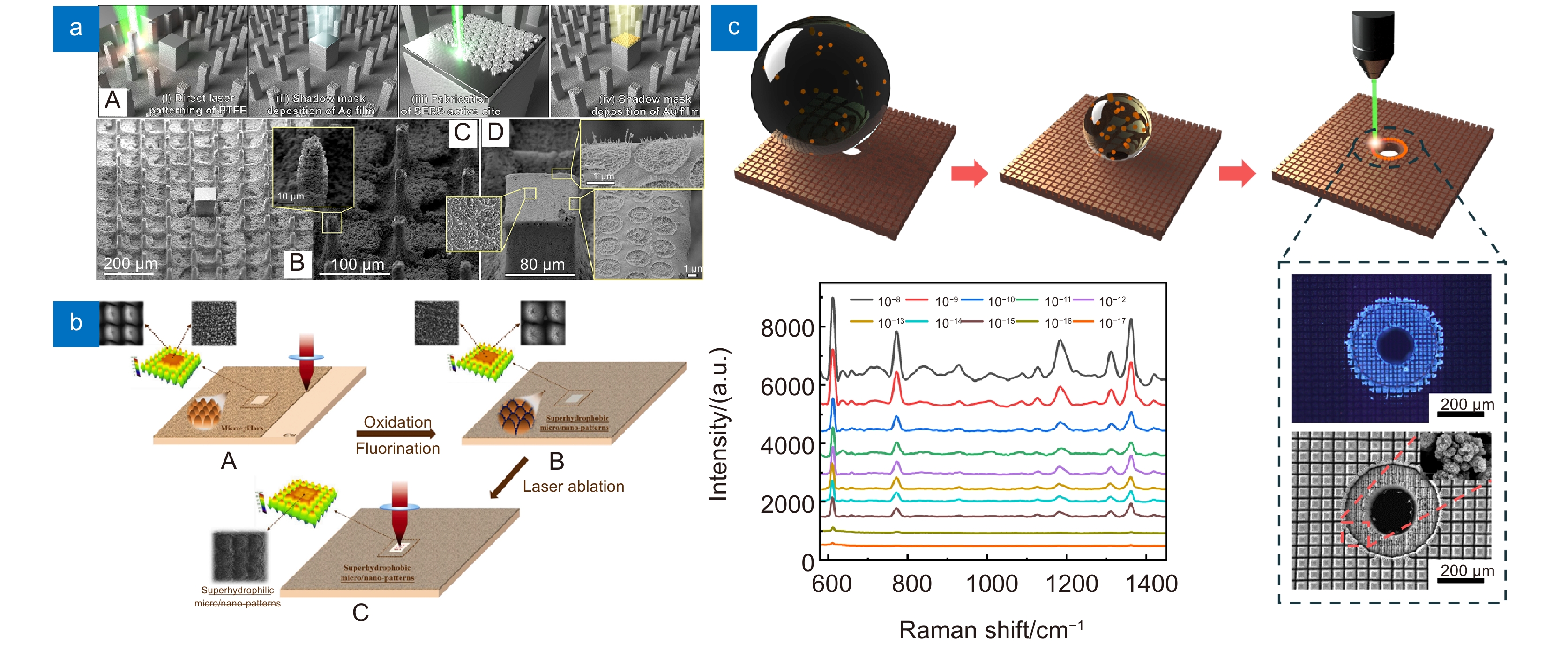
图 7 (a) 超敏感SERS基底的制造示意图 [74]; (b) 混合超亲水/超疏水基底的制造示意图 [75]; (c) 混合型超亲水/超疏水微孔SERS基底的示意图 [27]
Figure 7. (a) Schematic of the fabrication strategy of ultrasensitive SERS substrates [74]; (b) Schematic of the fabrication strategy of hybrid superhydrophilic/superhydrophobic substrates [75]; (c) Schematic of hybrid superhydrophilic/superhydrophobic microporous SERS substrates [27]
表 1 基于超快激光制备的各种类型SERS传感器的性能对比
Table 1. Performance comparison among various types of SERS sensors prepared by the ultrafast laser
基底材料 活性热点 加工方式 表面润湿性 分析物 检测极限/(mol/L) 增强因子 参考文献 活性热点的增加 微通道 银纳米颗粒 飞秒激光 / 对氨基苯硫酚 10−10 4 × 10⁸ [22] 微通道 银/钯合金纳米颗粒 飞秒激光 / R6G 10−9 2.62 × 10⁸ [31] 硅 银纳米颗粒 飞秒激光 亲水 R6G 10−12 / [33] 银 银纳米颗粒 飞秒激光 / R6G 10−8 1.2 × 10⁶ [42] 银 银纳米颗粒 飞秒激光 / R6G 10−8 5.6 × 10⁶ [44] 金 金纳米颗粒 飞秒激光 / DPA 10−15 / [47] 硅 金纳米颗粒 飞秒激光 / R6G / 4.3 × 10⁷ [62] 钛 银纳米颗粒 飞秒激光/水热法 / R6G 10−14 1.2 × 10⁹ [61] 铜 银纳米颗粒 纳秒激光/热氧化 / 4-甲苯硫酚 / 1.44 × 10⁵ [60] 目标分子的富集 硅 银纳米颗粒 飞秒激光 超疏水 R6G 10−14 6 × 10⁶ [72] 铜 铜纳米颗粒 飞秒激光 超疏水 R6G 10−13 1.2 × 10⁵ [71] 不锈钢 银纳米颗粒 飞秒激光 超疏水/疏水 R6G 10−14 5.7 × 10⁸ [68] 硅/PDMS 金纳米颗粒 飞秒激光 超疏水/超亲水 R6G 10−16 / [35] 聚四氟乙烯基 银纳米颗粒 飞秒激光 超疏水/超亲水 R6G 10−12 1 × 10⁷ [74] 铜 纳米金星 飞秒激光 超疏水/超亲水 R6G 10−18 1.09 × 10¹⁴ [75] 铜 银纳米颗粒 飞秒激光 超疏水/超亲水/微孔 R6G 10−17 5.19 × 10¹³ [27]  下载: 导出CSV
-
[1] Raman C V, Krishnan K S. A new type of secondary radiation[J]. Nature, 1928, 121(3048): 501−502. doi: 10.1038/121501c0
[2] Zhang X L, Dai Z G, Zhang X G, et al. Recent progress in the fabrication of SERS substrates based on the arrays of polystyrene nanospheres[J]. Sci China Phys Mech Astron, 2016, 59(12): 126801. doi: 10.1007/s11433-016-0341-y
[3] 陈娜. 硅基SERS芯片的构建及其在环境检测中的应用[D]. 苏州: 苏州大学, 2018.
Chen N. Construction of silicon-based SERS chips and their use in environmental detection[D]. Suzhou: Soochow University, 2018.
[4] 刘江涛, 洪昕. 基于微流控芯片SERS生物传感器的发展与应用[J]. 北京生物医学工程, 2018, 37(2): 201−207. doi: 10.3969/j.issn.1002-3208.2018.02.016
Liu J T, Hong X. Development and application of SERS biosensor based on microfluidic chip[J]. Beijing Biomed Eng, 2018, 37(2): 201−207. doi: 10.3969/j.issn.1002-3208.2018.02.016
[5] Wang X, Huang S C, Hu S, et al. Fundamental understanding and applications of Plasmon-enhanced Raman spectroscopy[J]. Nat Rev Phys, 2020, 2(5): 253−271. doi: 10.1038/s42254-020-0171-y
[6] Fleischmann M, Hendra P J, McQuillan A J. Raman spectra of pyridine adsorbed at a silver electrode[J]. Chem Phys Lett, 1974, 26(2): 163−166. doi: 10.1016/0009-2614(74)85388-1
[7] Jeanmaire D L, van Duyne R P. Surface Raman spectroelectrochemistry: part I. Heterocyclic, aromatic, and aliphatic amines adsorbed on the anodized silver electrode[J]. J Electroanal Chem Interfacial Electrochem, 1977, 84(1): 1−20. doi: 10.1016/S0022-0728(77)80224-6
[8] Chang T H P, Mankos M, Lee K Y, et al. Multiple electron-beam lithography[J]. Microelectron Eng, 2001, 57–58: 117−135. doi: 10.1016/S0167-9317(01)00528-7
[9] Garg V, Mote R G, Fu J. Focused ion beam direct fabrication of subwavelength nanostructures on silicon for multicolor generation[J]. Adv Mater Technol, 2018, 3(8): 1800100. doi: 10.1002/admt.201800100
[10] Chen W X, Tymchenko M, Gopalan P, et al. Large-area nanoimprinted colloidal Au nanocrystal-based nanoantennas for ultrathin polarizing plasmonic metasurfaces[J]. Nano Lett, 2015, 15(8): 5254−5260. doi: 10.1021/acs.nanolett.5b02647
[11] Vishnu J, Manivasagam V K, Gopal V, et al. Hydrothermal treatment of etched titanium: a potential surface Nano-modification technique for enhanced biocompatibility[J]. Nanomed-Nanotechnol Biol Med, 2019, 20: 102016. doi: 10.1016/j.nano.2019.102016
[12] Acharya G, Shin C S, McDermott M, et al. The hydrogel template method for fabrication of homogeneous Nano/microparticles[J]. J Control Release, 2010, 141(3): 314−319. doi: 10.1016/j.jconrel.2009.09.032
[13] Li H Z, Yang Q, Hou J, et al. Bioinspired micropatterned superhydrophilic Au-areoles for surface-enhanced Raman scattering (SERS) trace detection[J]. Adv Funct Mater, 2018, 28(21): 1800448. doi: 10.1002/adfm.201800448
[14] Agarwal P, Wang H, Sun M R, et al. Microfluidics enabled bottom-up engineering of 3D vascularized tumor for drug discovery[J]. ACS Nano, 2017, 11(7): 6691−6702. doi: 10.1021/acsnano.7b00824
[15] 廉洁, 周稳稳, 石西增, 等. 多靶标生物标志物检测的微流体磁敏生物传感器研制[J]. 分析化学, 2013, 41(9): 1302−1307. doi: 10.3724/SP.J.1096.2013.30231
Lian J, Zhou W W, Shi X Z, et al. Development of integrated microfluidic magnetic biosensor for multi-biomarker detection[J]. Chin J Anal Chem, 2013, 41(9): 1302−1307. doi: 10.3724/SP.J.1096.2013.30231
[16] Geissler M, Clime L, Hoa X D, et al. Microfluidic integration of a cloth-based hybridization array system (CHAS) for rapid, colorimetric detection of enterohemorrhagic Escherichia coli (EHEC) using an articulated, centrifugal platform[J]. Anal Chem, 2015, 87(20): 10565−10572. doi: 10.1021/acs.analchem.5b03085
[17] Liu X, Du D, Mourou G. Laser ablation and micromachining with ultrashort laser pulses[J]. IEEE J Quantum Electron, 1997, 33(10): 1706−1716. doi: 10.1109/3.631270
[18] 杨青, 成扬, 方政, 等. 仿生超滑表面的飞秒激光微纳制造及应用[J]. 光电工程, 2022, 49(1): 210326. doi: 10.12086/oee.2022.210326
Yang Q, Cheng Y, Fang Z, et al. The preparation and applications of bio-inspired slippery surface by femtosecond laser micro-Nano manufacturing[J]. Opto-Electron Eng, 2022, 49(1): 210326. doi: 10.12086/oee.2022.210326
[19] Shao Q, Que R H, Shao M W, et al. Copper nanoparticles grafted on a silicon wafer and their excellent surface-enhanced Raman scattering[J]. Adv Funct Mater, 2012, 22(10): 2067−2070. doi: 10.1002/adfm.201102943
[20] Liu Z, Cheng L, Zhang L, et al. Large-area fabrication of highly reproducible surface enhanced Raman substrate via a facile double sided tape-assisted transfer approach using hollow Au-Ag alloy nanourchins[J]. Nanoscale, 2014, 6(5): 2567−2572. doi: 10.1039/C3NR05840A
[21] Crouch C H, Carey J E, Warrender J M, et al. Comparison of structure and properties of femtosecond and nanosecond laser-structured silicon[J]. Appl Phys Lett, 2004, 84(11): 1850−1852. doi: 10.1063/1.1667004
[22] Xu B B, Ma Z C, Wang L, et al. Localized flexible integration of high-efficiency surface enhanced Raman scattering (SERS) monitors into microfluidic channels[J]. Lab Chip, 2011, 11(19): 3347−3351. doi: 10.1039/c1lc20397e
[23] Xu L M, Liu H G, Zhou H, et al. One-step fabrication of metal nanoparticles on polymer film by femtosecond LIPAA method for SERS detection[J]. Talanta, 2021, 228: 122204. doi: 10.1016/j.talanta.2021.122204
[24] de Angelis F, Gentile F, Mecarini F, et al. Breaking the diffusion limit with super-hydrophobic delivery of molecules to plasmonic nanofocusing SERS structures[J]. Nat Photonics, 2011, 5(11): 682−687. doi: 10.1038/nphoton.2011.222
[25] He A, Yang H, Xue W, et al. Tunable coffee-ring effect on a superhydrophobic surface[J]. Opt Lett, 2017, 42(19): 3936−3939. doi: 10.1364/OL.42.003936
[26] Song J, Cheng W F, Nie M T, et al. Partial leidenfrost evaporation-assisted ultrasensitive surface-enhanced Raman spectroscopy in a Janus water droplet on hierarchical plasmonic micro-/nanostructures[J]. ACS Nano, 2020, 14(8): 9521−9531. doi: 10.1021/acsnano.0c04239
[27] Yu J, Wu J G, Yang H, et al. Extremely sensitive SERS sensors based on a femtosecond laser-fabricated superhydrophobic/-philic microporous platform[J]. ACS Appl Mater Interfaces, 2022, 14(38): 43877−43885. doi: 10.1021/acsami.2c10381
[28] Chen S, Meng L Y, Shan H Y, et al. How to light special hot spots in Multiparticle-film configurations[J]. ACS Nano, 2016, 10(1): 581−587. doi: 10.1021/acsnano.5b05605
[29] Willets K A, van Duyne R P. Localized surface Plasmon resonance spectroscopy and sensing[J]. Annu Rev Phys Chem, 2007, 58: 267−297. doi: 10.1146/annurev.physchem.58.032806.104607
[30] Di Pietro P, Strano G, Zuccarello L, et al. Gold and silver nanoparticles for applications in theranostics[J]. Curr Top Med Chem, 2016, 16(27): 3069−3102. doi: 10.2174/1568026616666160715163346
[31] Ma Z C, Zhang Y L, Han B, et al. Femtosecond laser direct writing of plasmonic Ag/Pd alloy nanostructures enables flexible integration of robust SERS substrates[J]. Adv Mater Technol, 2017, 2(6): 1600270. doi: 10.1002/admt.201600270
[32] Yan W J, Yang L K, Chen J N, et al. In situ two-step Photoreduced SERS materials for on-chip single-molecule spectroscopy with high reproducibility[J]. Adv Mater, 2017, 29(36): 1702893. doi: 10.1002/adma.201702893
[33] Ran P, Jiang L, Li X, et al. Femtosecond photon-mediated plasma enhances photosynthesis of plasmonic nanostructures and their SERS applications[J]. Small, 2019, 15(11): 1804899. doi: 10.1002/smll.201804899
[34] Delaporte P, Alloncle A P. Laser-induced forward transfer: a high resolution additive manufacturing technology[J]. Opt Laser Technol, 2016, 78: 33−41. doi: 10.1016/j.optlastec.2015.09.022
[35] Ma X D, Jiang L, Li X W, et al. Hybrid Superhydrophilic-superhydrophobic micro/nanostructures fabricated by femtosecond laser-induced forward transfer for sub-femtomolar Raman detection[J]. Microsyst Nanoeng, 2019, 5: 48. doi: 10.1038/s41378-019-0090-1
[36] Jing Y T, Wang R J, Wang Q L, et al. An overview of surface-enhanced Raman scattering substrates by pulsed laser deposition technique: fundamentals and applications[J]. Adv Compos Hybrid Mater, 2021, 4(4): 885−905. doi: 10.1007/s42114-021-00330-0
[37] Khan T M, Lunney J G, O'Rourke D, et al. Various pulsed laser deposition methods for preparation of silver-sensitised glass and paper substrates for surface-enhanced Raman spectroscopy[J]. Appl Phys A, 2019, 125(9): 659. doi: 10.1007/s00339-019-2968-z
[38] Smyth C A, Mirza I, Lunney J G, et al. Surface-enhanced Raman spectroscopy (SERS) using Ag nanoparticle films produced by pulsed laser deposition[J]. Appl Surf Sci, 2013, 264: 31−35. doi: 10.1016/j.apsusc.2012.09.078
[39] 杨焕, 曹宇, 李峰平, 等. 激光制备超疏水表面研究进展[J]. 光电工程, 2017, 44(12): 1160−1168. doi: 10.3969/j.issn.1003-501X.2017.12.003
Yang H, Cao Y, Li F P, et al. Research progress in superhydrophobic surfaces fabricated by laser[J]. Opto-Electron Eng, 2017, 44(12): 1160−1168. doi: 10.3969/j.issn.1003-501X.2017.12.003
[40] 欧阳旭, 谢子健, 张孟瑞, 等. 基于激光诱导表面周期结构的微纳防伪结构色[J]. 光电工程, 2022, 49(1): 210320. doi: 10.12086/oee.2022.210320
Ouyang X, Xie Z J, Zhang M R, et al. Laser-induced periodic surface structure for microscale anti-counterfeiting structural colors[J]. Opto-Electron Eng, 2022, 49(1): 210320. doi: 10.12086/oee.2022.210320
[41] 朱小伟, 潘哲豪, 杨文锋, 等. 基于激光三维雕刻的CFRP多梯层挖补胶接接头加工技术研究[J]. 光电工程, 2022, 49(1): 210314. doi: 10.12086/oee.2022.210314
Zhu X W, Pan Z H, Yang W F, et al. Study on multi-layered CFRP patch bonding joint based on laser 3D engraving technology[J]. Opto-Electron Eng, 2022, 49(1): 210314. doi: 10.12086/oee.2022.210314
[42] Chang H W, Tsai Y C, Cheng C W, et al. Nanostructured Ag surface fabricated by femtosecond laser for surface-enhanced Raman scattering[J]. J Colloid Interface Sci, 2011, 360(1): 305−308. doi: 10.1016/j.jcis.2011.04.005
[43] Erol M, Han Y, Stanley S K, et al. SERS not to be taken for granted in the presence of oxygen[J]. J Am Chem Soc, 2009, 131(22): 7480−7481. doi: 10.1021/ja807458x
[44] Luo X, Liu W J, Chen C H, et al. Femtosecond laser micro-Nano structured Ag SERS substrates with unique sensitivity, uniformity and stability for food safety evaluation[J]. Opt Laser Technol, 2021, 139: 106969. doi: 10.1016/j.optlastec.2021.106969
[45] Siddhanta S, Thakur V, Narayana C, et al. Universal metal-semiconductor hybrid nanostructured SERS substrate for biosensing[J]. ACS Appl Mater Interfaces, 2012, 4(11): 5807−5812. doi: 10.1021/am302102p
[46] Fan M K, Andrade G F S, Brolo A G. A review on the fabrication of substrates for surface enhanced Raman spectroscopy and their applications in analytical chemistry[J]. Anal Chim Acta, 2011, 693(1–2): 7−25. doi: 10.1016/j.aca.2011.03.002
[47] Naqvi T K, Bajpai A, Bharati M S S, et al. Ultra-sensitive reusable SERS sensor for multiple hazardous materials detection on single platform[J]. J Hazard Mater, 2021, 407: 124353. doi: 10.1016/j.jhazmat.2020.124353
[48] Xu K C, Wang Z Y, Tan C F, et al. Uniaxially stretched flexible surface Plasmon resonance film for versatile surface enhanced Raman scattering diagnostics[J]. ACS Appl Mater Interfaces, 2017, 9(31): 26341−26349. doi: 10.1021/acsami.7b06669
[49] Dai H C, Sun Y J, Ni P J, et al. Three-dimensional TiO2 supported silver nanoparticles as sensitive and UV-cleanable substrate for surface enhanced Raman scattering[J]. Sens Actuators B Chem, 2017, 242: 260−268. doi: 10.1016/j.snb.2016.10.085
[50] Xu K C, Zhou R, Takei K, et al. Toward flexible surface-enhanced Raman scattering (SERS) sensors for point-of-care diagnostics[J]. Adv Sci, 2019, 6(16): 1900925. doi: 10.1002/advs.201900925
[51] Zhang Q, Lee Y H, Phang I Y, et al. Hierarchical 3D SERS substrates fabricated by integrating photolithographic microstructures and self-assembly of silver nanoparticles[J]. Small, 2014, 10(13): 2703−2711. doi: 10.1002/smll.201303773
[52] Phan-Quang G C, Han X M, Koh C S L, et al. Three-dimensional surface-enhanced Raman scattering platforms: large-scale plasmonic hotspots for new applications in sensing, microreaction, and data storage[J]. Acc Chem Res, 2019, 52(7): 1844−1854. doi: 10.1021/acs.accounts.9b00163
[53] Yang D, Cho H, Koo S, et al. Simple, large-scale fabrication of uniform Raman-enhancing substrate with enhancement saturation[J]. ACS Appl Mater Interfaces, 2017, 9(22): 19092−19101. doi: 10.1021/acsami.7b03239
[54] Li X, Lee H K, Phang I Y, et al. Superhydrophobic-oleophobic Ag nanowire platform: an analyte-concentrating and quantitative aqueous and organic toxin surface-enhanced Raman scattering sensor[J]. Anal Chem, 2014, 86(20): 10437−10444. doi: 10.1021/ac502955w
[55] Lee Y H, Lay C L, Shi W X, et al. Creating two self-assembly micro-environments to achieve supercrystals with dual structures using polyhedral nanoparticles[J]. Nat Commun, 2018, 9(1): 2769. doi: 10.1038/s41467-018-05102-x
[56] Tao A, Kim F, Hess C, et al. Langmuir-Blodgett silver nanowire monolayers for molecular sensing using surface-enhanced Raman spectroscopy[J]. Nano Lett, 2003, 3(9): 1229−1233. doi: 10.1021/nl0344209
[57] Khew S Y, Tan C F, Yan H P, et al. Nanosecond laser ablation for enhanced adhesion of CuO nanowires on copper substrate and its application for oil-water separation[J]. Appl Surf Sci, 2019, 465: 995−1002. doi: 10.1016/j.apsusc.2018.09.256
[58] Zhao Y Z, Su Y L, Hou X Y, et al. Directional sliding of water: biomimetic snake scale surfaces[J]. Opto-Electron Adv, 2021, 4(4): 210008. doi: 10.29026/oea.2021.210008
[59] Lin Z Y, Hong M H. Femtosecond laser precision engineering: from micron, submicron, to nanoscale[J]. Ultrafast Sci, 2021, 2021: 9783514. doi: 10.34133/2021/9783514
[60] Xu K C, Yan H P, Tan C F, et al. Hedgehog inspired CuO nanowires/Cu2O composites for broadband visible-light-driven recyclable surface enhanced Raman scattering[J]. Adv Opt Mater, 2018, 6(7): 1701167. doi: 10.1002/adom.201701167
[61] Lu J L, Yang J J, Singh S C, et al. Hierarchical micro/nanostructured TiO2/Ag substrates based on femtosecond laser structuring: a facile route for enhanced SERS performance and location predictability[J]. Appl Surf Sci, 2019, 478: 737−743. doi: 10.1016/j.apsusc.2019.01.257
[62] Li C, Hu J, Jiang L, et al. Shaped femtosecond laser induced photoreduction for highly controllable Au nanoparticles based on localized field enhancement and their SERS applications[J]. Nanophotonics, 2020, 9(3): 691−702. doi: 10.1515/nanoph-2019-0460
[63] Sheehan P E, Whitman L J. Detection limits for nanoscale biosensors[J]. Nano Lett, 2005, 5(4): 803−807. doi: 10.1021/nl050298x
[64] Wang W D, Yin Y G, Tan Z Q, et al. Coffee-ring effect-based simultaneous SERS substrate fabrication and analyte enrichment for trace analysis[J]. Nanoscale, 2014, 6(16): 9588−9593. doi: 10.1039/C4NR03198A
[65] Ji B, Zhang L J, Li M Z, et al. Suppression of coffee-ring effect via periodic oscillation of substrate for ultra-sensitive enrichment towards surface-enhanced Raman scattering[J]. Nanoscale, 2019, 11(43): 20534−20545. doi: 10.1039/C9NR06989E
[66] Fang Y, Seong N H, Dlott D D. Measurement of the distribution of site enhancements in surface-enhanced Raman scattering[J]. Science, 2008, 321(5887): 388−392. doi: 10.1126/science.1159499
[67] Gentile F, Das G, Coluccio M L, et al. Ultra low concentrated molecular detection using super hydrophobic surface based biophotonic devices[J]. Microelectron Eng, 2010, 87(5–8): 798−801. doi: 10.1016/j.mee.2009.11.083
[68] Yang H, Guan X Y, Pang G, et al. Femtosecond laser patterned superhydrophobic/hydrophobic SERS sensors for rapid positioning ultratrace detection[J]. Opt Express, 2021, 29(11): 16904−16913. doi: 10.1364/OE.423789
[69] Xu F G, Zhang Y, Sun Y J, et al. Silver nanoparticles coated zinc oxide nanorods array as superhydrophobic substrate for the amplified SERS effect[J]. J Phys Chem C, 2011, 115(20): 9977−9983. doi: 10.1021/jp201897j
[70] Wallace R A, Charlton J J, Kirchner T B, et al. Superhydrophobic analyte concentration utilizing colloid-pillar array SERS substrates[J]. Anal Chem, 2014, 86(23): 11819−11825. doi: 10.1021/ac5033947
[71] Fu P, Shi X S, Jiang F, et al. Superhydrophobic nanostructured copper substrate as sensitive SERS platform prepared by femtosecond laser pulses[J]. Appl Surf Sci, 2020, 501: 144269. doi: 10.1016/j.apsusc.2019.144269
[72] Wang A D, Jiang L, Li X W, et al. Low-adhesive superhydrophobic surface-enhanced Raman spectroscopy substrate fabricated by femtosecond laser ablation for ultratrace molecular detection[J]. J Mater Chem B, 2017, 5(4): 777−784. doi: 10.1039/C6TB02629J
[73] Huang J B, Wen Y H, Li J, et al. Superhydrophobic-superhydrophilic hybrid surface with highly ordered tip-capped nanopore arrays for surface-enhanced Raman scattering spectroscopy[J]. ACS Appl Mater Interfaces, 2020, 12(33): 37499−37505. doi: 10.1021/acsami.0c12127
[74] Pavliuk G, Pavlov D, Mitsai E, et al. Ultrasensitive SERS-based plasmonic sensor with analyte enrichment system produced by direct laser writing[J]. Nanomaterials, 2020, 10(1): 49. doi: 10.3390/nano10010049
[75] Luo X, Pan R, Cai M Y, et al. Atto-molar Raman detection on patterned superhydrophilic-superhydrophobic platform via localizable evaporation enrichment[J]. Sens Actuators B Chem, 2021, 326: 128826. doi: 10.1016/j.snb.2020.128826
[76] Patil N D, Bange P G, Bhardwaj R, et al. Effects of substrate heating and wettability on evaporation dynamics and deposition patterns for a sessile water droplet containing colloidal particles[J]. Langmuir, 2016, 32(45): 11958−11972. doi: 10.1021/acs.langmuir.6b02769
[77] Chen Y X, Xu L P, Meng J X, et al. Superwettable microchips with improved spot homogeneity toward sensitive biosensing[J]. Biosens Bioelectron, 2018, 102: 418−424. doi: 10.1016/j.bios.2017.11.036
-
 点击扫一扫
点击扫一扫
图(7)
表(1)
计量
- 文章访问数: 6470
- PDF下载数: 1617
- 施引文献: 0


