
 E-mail Alert
 RSS
RSS
Research progress in the quantum dots of nonmetals and their compounds prepared by ultrasonic method
-
摘要
量子点(Quantum dots,QDs)晶粒直径在1~20 nm之间,是一种零维的纳米材料,具有尺寸可调、激发光谱宽、高量子效率、宽波长范围、光化学稳定性高、不易光解等优异的光学特性。当量子点的尺寸接近或小于激子波尔半径时,原有的连续能带结构量子化,其性质也发生显著变化,表现出优异的光电性能。本综述介绍了量子点的制备方法,其中,超声法作为一种常见的“自上而下”方法,因操作简单、对环境友好等优点被广泛使用。首先,概述了利用超声法制备非金属和非金属化合物量子点的机理和表征技术,并分析了分散剂、超声功率和时间等因素对其尺寸及形貌的影响。随后,对超声法制备的非金属和非金属化合物量子点在激光器、太阳能电池等领域的应用展开了讨论,并针对目前研究存在的一些挑战和难题,提出了自己的观点和见解。最后,对超声法制备非金属单质及非金属化合物量子点进行了总结和展望。
Abstract
Quantum dots (QDs), with diameters ranging from 1 to 20 nm, are zero-dimensional nanomaterials. They possess excellent optical properties, including size tunability, broad excitation spectra, high quantum efficiency, wide wavelength range, high photostability, and low photolysis. When the size of QDs approaches or becomes smaller than the exciton Bohr radius, the original material's continuous band structure undergoes quantization, leading to significant changes in properties and exhibiting outstanding optoelectronics performance. This review introduces the preparation methods of QDs, among which the ultrasonic method, as a common "top-down" method, is widely used because of its advantages of simple operation and environmental friendliness. Firstly, the mechanism and characterization techniques of non-metal and non-metallic compound QDs prepared by the ultrasonic method were prepared, and the effects of dispersants agent, ultrasonic power, and time on their size and morphology were analyzed. Subsequently, the application of non-metal and non-metallic compounds QDs prepared by the ultrasonic method in laser, solar cell, and other fields is discussed. The challenges and issues in current research are addressed, and personal perspectives and insights are provided. Finally, the prospect is given.
-
Key words:
- quantum dots /
- nanomaterials /
- ultrasonic method /
- non-metal /
- non-metallic compound
-
Overview
Overview: This review paper aims to provide a detailed introduction and summary of the research progress on the preparation of non-metal quantum dots (QDs) using the ultrasonic method, and explore their potential applications in various fields. QDs are nanomaterials with zero-dimensional structures, with grain diameters ranging from 1-20 nm. Compared to traditional materials, QDs exhibit a wide excitation spectrum, continuous distribution characteristics, symmetric and narrow emission spectra, tunable colors, high photostability, and resistance to photobleaching, making them highly attractive for applications in optoelectronic devices, solar cells, optical devices, sensors, and bioimaging. The paper first introduces the preparation methods of QDs, among which the ultrasonic method is a common "top-down" approach known for its simplicity and environmental friendliness. When the size of QDs approaches or is smaller than the exciton Bohr radius, the continuous band structure of the original material becomes quantized, resulting in significant changes in their properties. Subsequently, an overview of the research progress in the preparation of non-metal quantum dots using the ultrasonic method is presented, including the preparation methods and characterization techniques for different non-metal and non-metallic compound quantum dots. During the preparation process, the action of ultrasound, which involves the formation, growth, and collapse of bubbles, accompanied by intense shock waves, can produce small-sized nanoscale particles.
Through a review and analysis of related studies, the following conclusions are drawn: the ultrasonic method is an effective approach for the preparation of non-metal quantum dots, offering advantages such as simplicity, low cost, controllable size, environmental friendliness, and scalability. However, there are still challenges in current research, such as controlling the size and morphology of QDs and improving their luminescence efficiency and stability. Therefore, by optimizing the preparation process of the ultrasonic method, the stability and dispersibility of QDs can be further improved, facilitating their in-depth research and application in the field of nanomaterials.
In summary, the preparation of non-metal quantum dots using the ultrasonic method is a research area with potential and challenges. Through continuous research and exploration, along with the development of new materials, the application of new processes, and interdisciplinary collaborations, the ultrasonic method for QD preparation will have broader prospects, providing new opportunities and breakthroughs for the development of optoelectronics, energy, and biomedical fields.
-

-
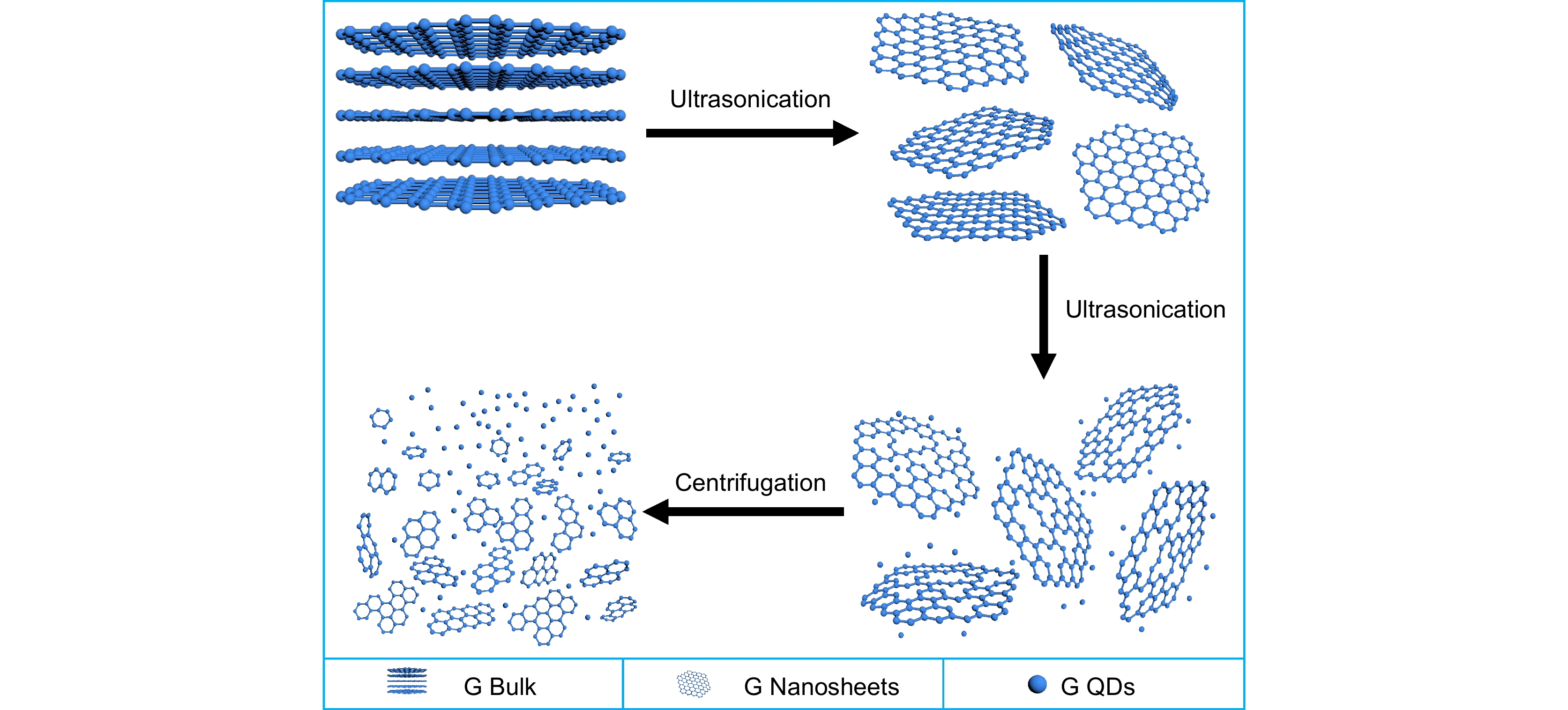
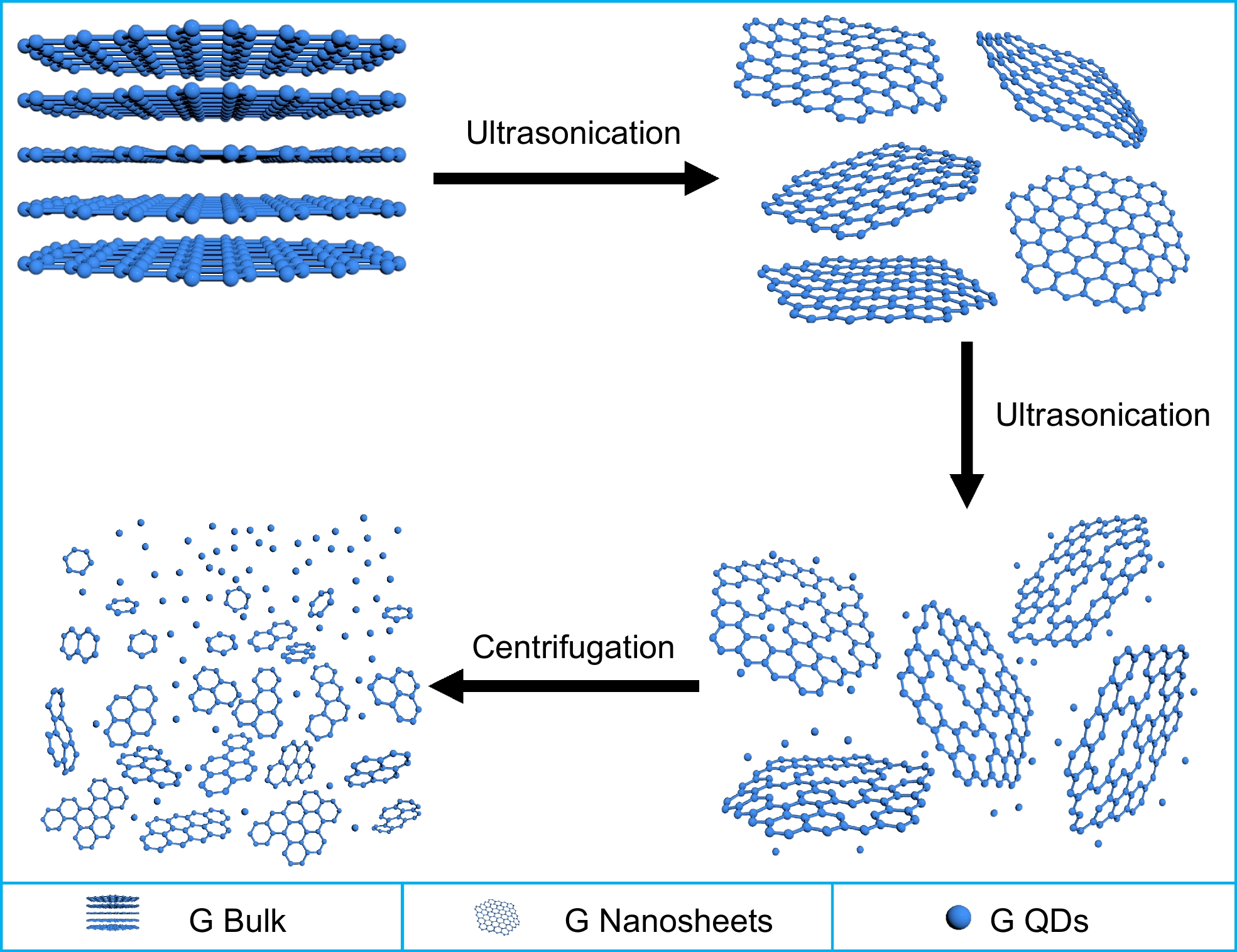
图 1 超声法制备G QDs的机理图
Figure 1. Mechanism diagram of G QDs prepared by the ultrasonic method
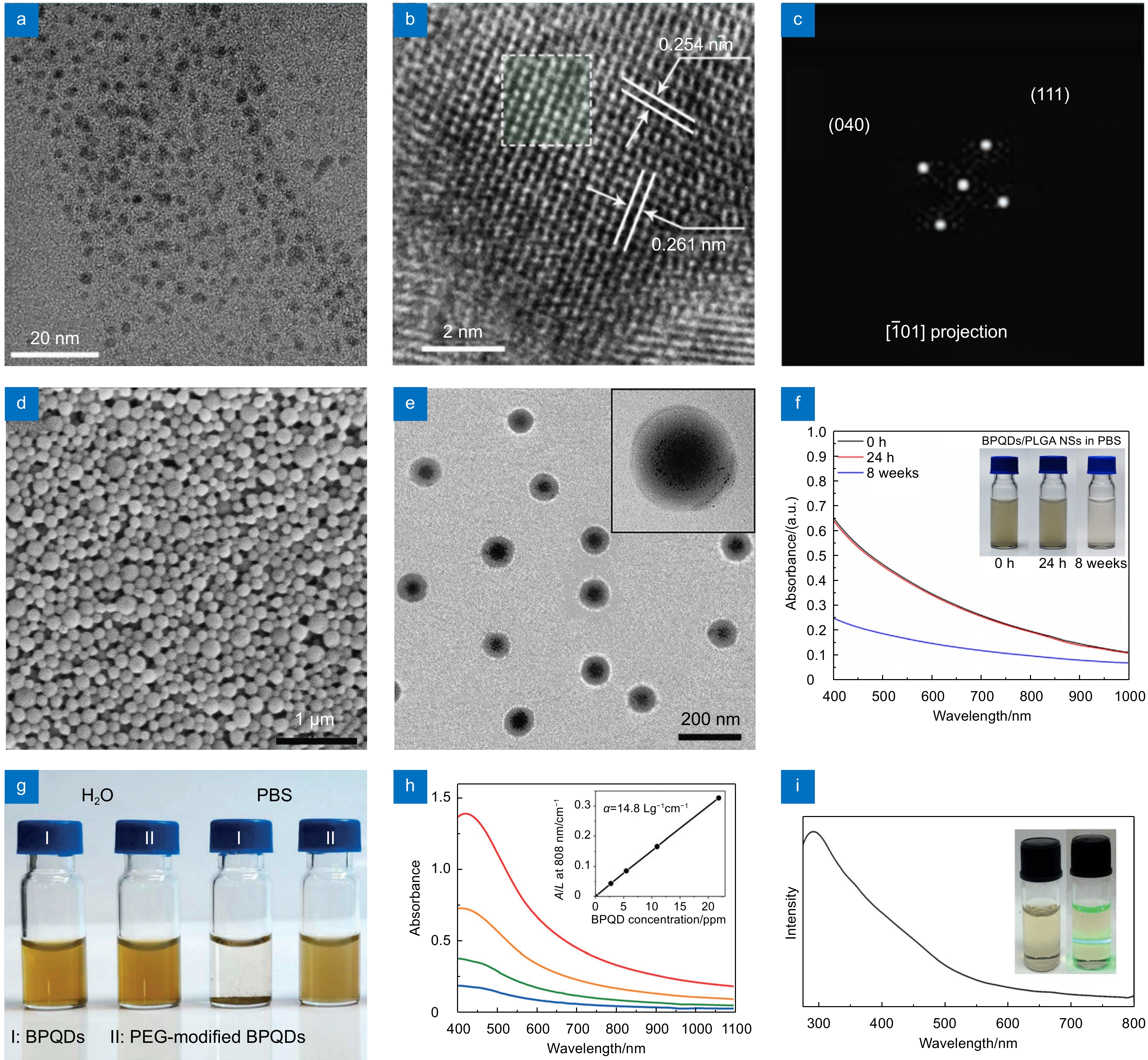
图 2 黑磷量子点的制备及表征。(a)和(b)分别为BP QD的TEM图和HRTEM图[62]; (c)为(b)中白色高亮部分的FFT图[62];(d)、(e)和(f)分别为BP QDs/PLGA NSs的SEM图、TEM图和在PBS中分散0 h、24 h和8周的吸收光谱[32];(g)和(h)分别为水或PBS溶液中的BP QDs (Ⅰ)与PEG修饰(Ⅱ)的照片和不同浓度的PEG修饰的BP QDs分散在水溶液中的吸收光谱[37];(i) BP QDs的紫外-可见吸收光谱[65]
Figure 2. Preparation and characterisation of BP QDs. (a) and (b) TEM and HRTEM images of the BP QD, respectively [62]; (c) FFT pattern of the white highlights in (b) [62]; (d) , (e) and (f) are the SEM and TEM images of BP QDs/PLGA NSs and the absorption spectra dispersed in PBS for 0 h, 24 h and 8 weeks, respectively[32]; (g) and (h) are the pictures of BP QDs (Ⅰ) and PEG modified (Ⅱ) in water or PBS solution, respectively, and the absorption spectra of different concentrations of PEG modified BP QDs dispersed in water solution[37]; (i) Ultraviolet visible absorption spectrum of BP QDs[65]
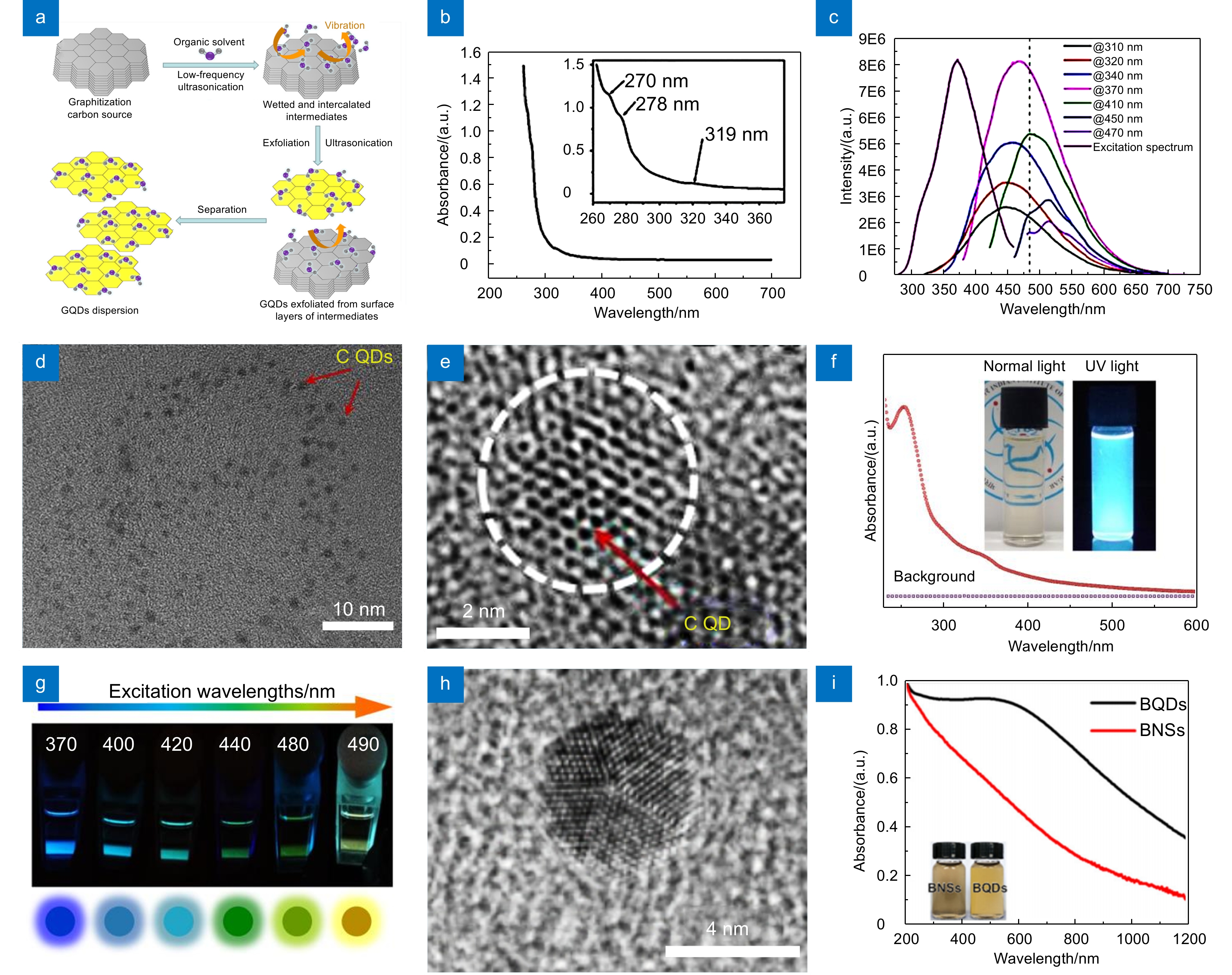
图 3 元素半导体量子点制备及表征。(a)超声剥离制备G QDs示意图[50];(b) G QDs的紫外-可见吸收光谱图[45];(c)在不同激发波长下的光致发光光谱以及激发光谱图[45];(d)和(e)分别为C QDs的TEM图和HRTEM图[35];(f)以非超声乙腈为溶剂背景的C QDs紫外-可见吸收光谱[35];(g)在不同的激发波长下激发的C QDs荧光的照片[35];(h)和(i)分别为B QDs的HRTEM图和紫外-可见吸收光谱图[44]
Figure 3. Preparation and characterization of elemental semiconductor QDs. (a) Schematic diagram of G QDs prepared by ultrasonic stripping [50]; (b) Ultraviolet-visible absorption spectra of G QDs[45]; (c) Photoluminescence spectra and excitation spectra at different excitation wavelengths[45]; (d) and (e) are TEM and HRTEM images of C QDs, respectively[35]; (f) The UV-visible absorption spectrum of CQDs dispersion along with non-sonicated acetonitrile as solvent background[35]; (g) Photographs of C QDs fluorescence excited at different excitation wavelengths[35]; (h) and (i) are HRTEM image and UV-vis absorption spectra of B QDs, respectively[44]
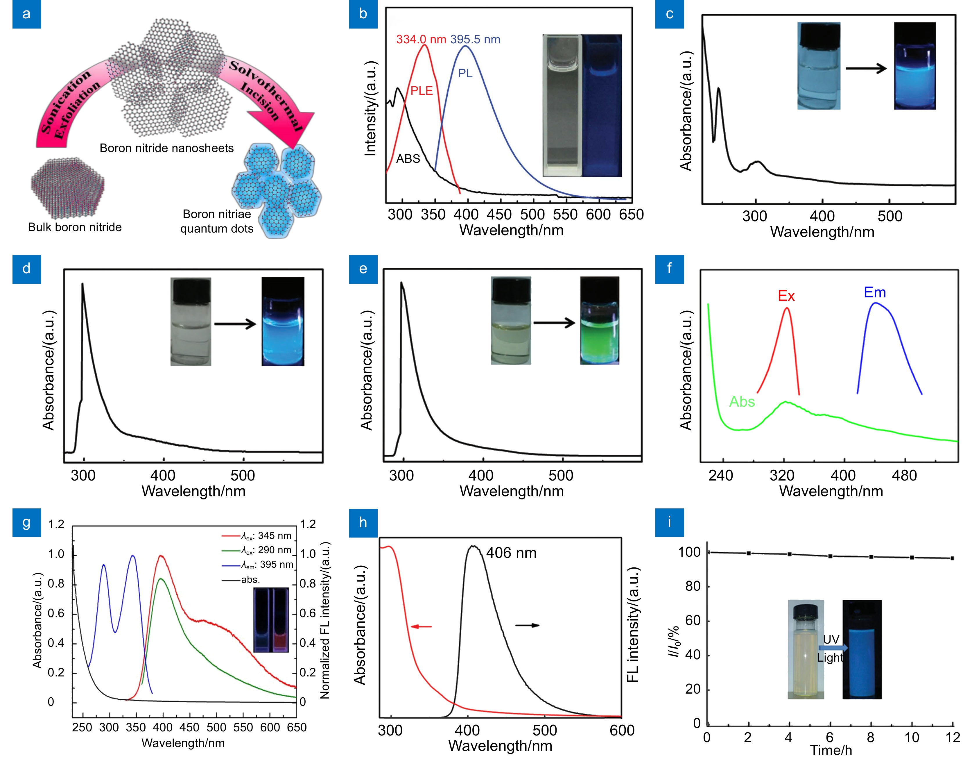
图 4 非金属化合物量子点的制备及表征。(a) BN QDs的制备示意图[73];(b) BN QDs的紫外-可见吸收 (黑色)、PLE (红色)和PL (蓝色)光谱图[71];(c)、(d)和(e)分别为分散在乙醇、DMF、NMP机溶剂的紫外-可见吸收光谱图[70];(f) OCNQDs的紫外-可见吸收和发射光谱图[75];(g) BCNO QDs的紫外-可见吸收光谱图[76];(h)和(i)分别为g-C3N4 QDs的紫外-可见光谱和PL强度与紫外光照明时间的关系[77]
Figure 4. Preparation and characterization of nonmetallic compounds QDs. (a) Schematic diagram of BN QDs preparation[73]; (b) Ultraviolet-visible absorption (black), PLE (red) and PL (blue) spectra of BN QDs[71]; (c), (d) and (e) are the ultraviolet-visible absorption spectra of organic solvents dispersed in ethanol, DMF, and NMP, respectively[70]; (f) Ultraviolet-visible absorption and emission spectra of OCNQDs[75]; (g) Ultraviolet-visible absorption spectra of BCNO QDs[76]; (h) and (i) are the Ultraviolet-visible spectra of g-C3N4 QDs and the relationship between PL intensity and UV illumination time, respectively[77]
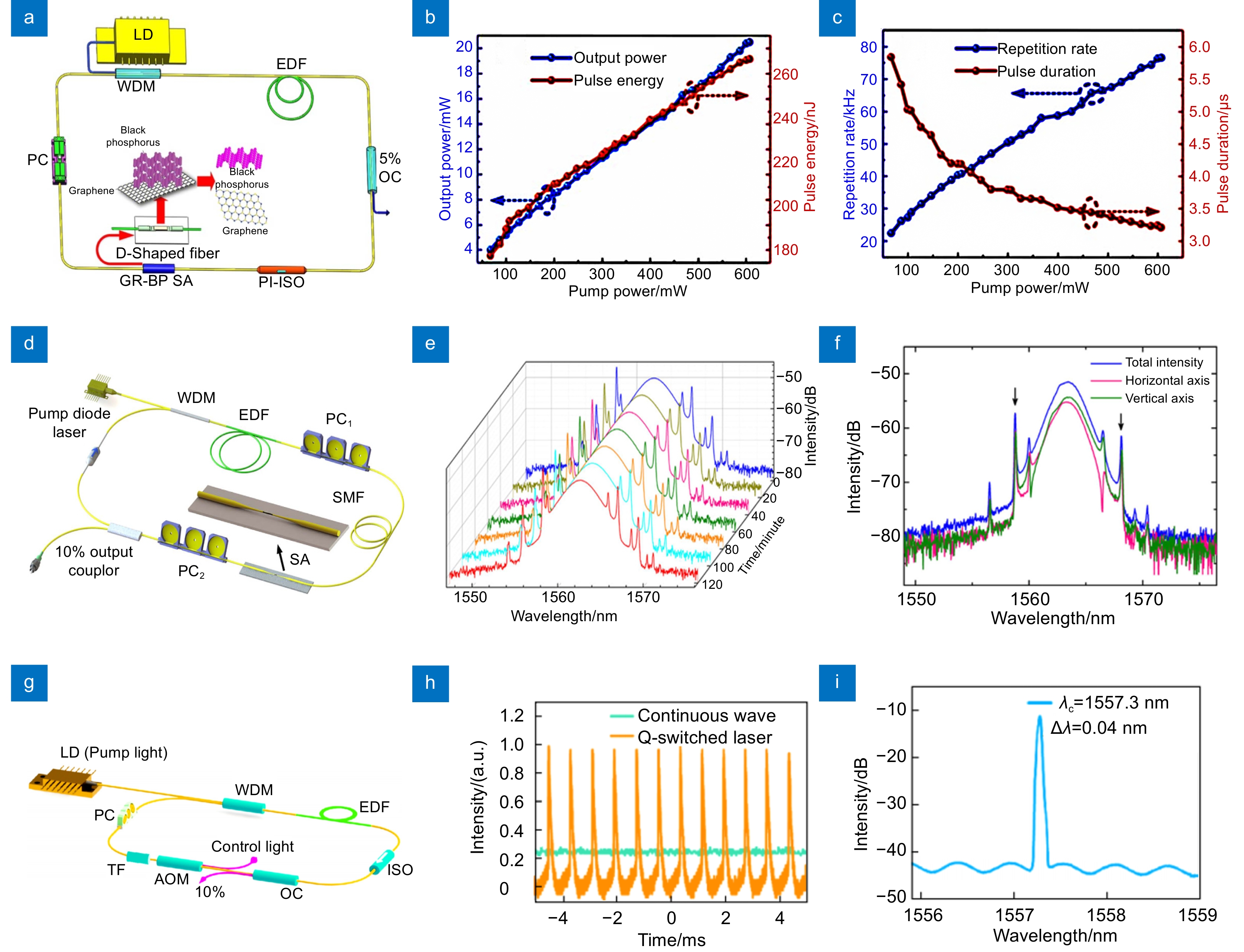
图 5 量子点激光器应用。(a)基于GR-BP的脉冲激光或超短脉冲激光[55];(b)和(c)分别为输出功率与脉冲能量作为泵功率函数和输出重复率与脉冲持续时间作为泵浦功率函数[55];(d)基于微纤维的P QD-SA的超快掺铒光纤激光器的示意图[47];(e)和(f)分别为光学光谱和孤子光谱[47];(g)基于B QDs的全光主动Q开关激光器的实验图[61];(h)和(i)分别为连续波与调Q脉冲激光器的状态图和输出光谱图[61]
Figure 5. Applications of QDs lasers. (a) Configuration of the pulsed laser or ultrashort pulsed laser based on GR-BP[55]; (b) and (c) are output power and pulse energy as pump power functions, and output repetition rate and pulse duration as pump power functions, respectively[55]; (d) Schematic diagram of ultrafast Erbium-doped fiber laser based on microfiber P QD-SA[47]; (e) and (f) are optical spectra and soliton spectra, respectively[47]; (g) Experimental diagram of all-optical active Q-switched lasers based on B QDs[61]; (h) and (i) are the state and output spectra of CW and Q-switched pulse lasers, respectively[61]
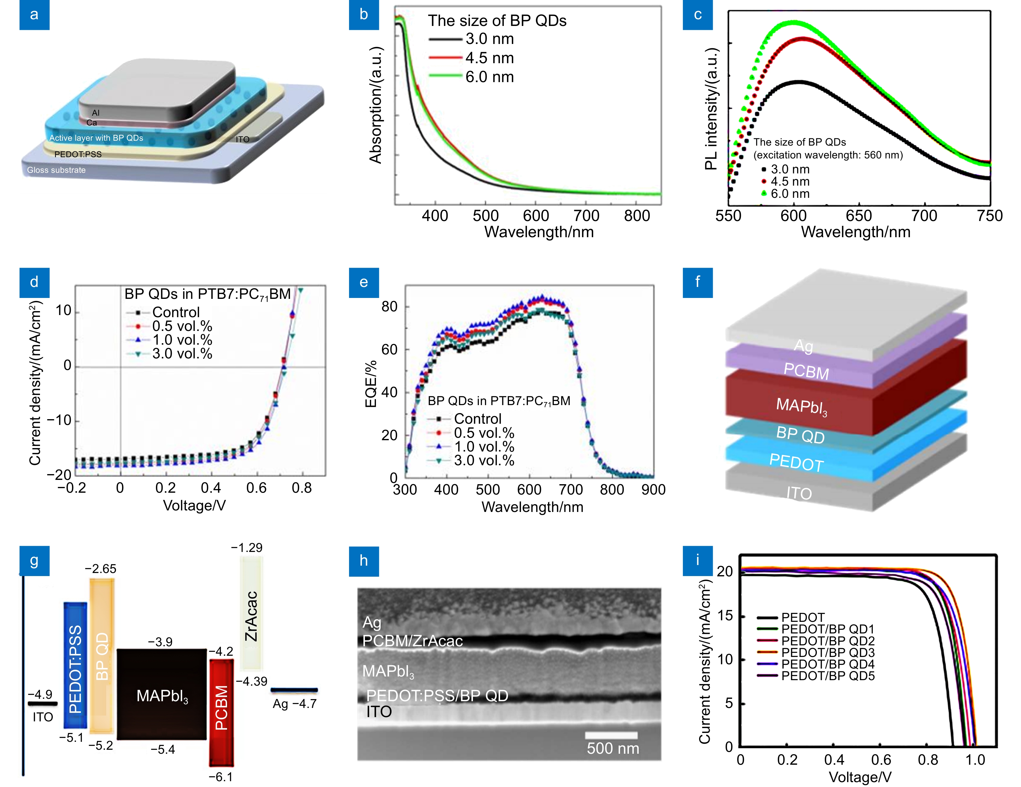
图 6 量子点光电器件应用。(a)以ITO玻璃为衬底的OPV器件结构[63];(b)和(c)分别为BP QDs在不同尺寸条件下的紫外-可见吸收光谱和PL光谱[63];(d)和(e)分别为不同浓度BP QD的OPV的 J-V特性曲线和EQEs测量[63];(f)和(g)分别为p-i-n平面HPSC的器件结构示意图及其器件中各层的能级图[64];(h) HPSCs结构的SEM图[64];(i)有和无BP QDs夹层的HPSCs的J-V特性曲线[64]
Figure 6. Optoelectronic device applications of QDs. (a) OPV device structure on the ITO glass substrate[63]; (b) and (c) are the UV visible absorption spectra and PL spectra of BP QDs under different size conditions, respectively[63]; (d) and (e) are respectively the J-V characteristic curves and EQEs measurements of OPVs with different concentrations of BP QD[63]; (f) and (g) are respectively the device structure diagram of p-i-n plane HPSC and the energy level diagram of each layer in the device[64]; (h) SEM diagram of the HPSCs structure[64]; (i) J-V characteristic curve of HPSCs with and without BP QDs interlayer[64]
表 1 超声法制备元素半导体量子点
Table 1. Preparation of elemental semiconductor QDs by the ultrasonic method
Materials Dispersants Ultrasonic conditions Size/nm Absorption
wavelength/nmApplication Year Ref. BP QDs NMP 100 W/7 h 2 - Electrocatalysts 2019 [53] 30 h 4.25 - Heterojunctions 2021 [46] 10 h 3.4 350~1000 Nonlinear optics 2016 [42] 6 h 3.3 - Lubrication 2022 [49] 200 W/3 h 4.9 300~800 Memory performance 2015 [65] 1200 W/3 h (Probe sonication)
300 W/10 h (Bath sonication)2.6 400~1100 Targeted photothermal cancer therapy 2015 [37] 1200 W/4 h (Probe sonication)
300 W/12 h (Bath sonication)3.1 400~1000 Photothermal cancer therapy 2016 [32] 200 W/3 h 2.6 - Saturable absorber device 2017 [47] 1200 W/3 h (Probe sonication)
5 h (Bath sonication)3.8 250~850 Ultra-broadband saturable absorbers 2017 [36] 600 W/6 h (Probe sonication)
300 W/10 h (Bath sonication)2.5 400~1000 Cancer treatment 2017 [48] 6 h 2.7 - Resistive random access memories 2017 [62] 1200 W/3 h (Probe sonication)
300 W/11 h (Bath sonication)2.6 - Biomedical research 2017 [67] 200 W/15 h 2.7 200~1000 Biological cells 2017 [66] 150 W/4 h 5.7 300~1500 Ultrafast photonics devices 2017 [60] 1200 W/3 h (Probe sonication)
300 W/10 h (Bath sonication)2.8 400~900 Efficient PA agents 2017 [39] Ethanol - 2.5 200~800 - 2020 [54] IPA 500 W/3 h 5.2 250~800 Electron-extraction layers 2017 [64] 40 kHz/2 h 2.82-10 - - 2020 [59] CF 1 h 5.9 240~800 Cellular imaging 2017 [52] NVP 20 W/5 h/ 5 ℃ 5 - FET devices 2016 [40] DI 100 W/30 min 10 - HeLa cell imaging 2016 [68] C QDs NMP 45 W/1 h 3 200~500 Fluorescence nanoprobes 2016 [50] 53 kHz/10 h 3.25 190~490 UV detector 2017 [57] 50 W/9 h 5.8 260~700 - 2018 [45] 10 h 3-5 - Sensors 2018 [33] DMSO 400 W/3 h 10-30 - - 2014 [56] H2SO4+HNO3 300 W/12 h 3-5 250~450 Photocatalyst 2012 [34] H2SO4+H2O2 120 W/24 h 4.45 200~800 Fluorescent sensor 2020 [41] Acetonitrile 500 W/4 h 2-3 250~600 - 2019 [35] Se QDs NMP 500 W/4 h 2.95 200~750 - 2017 [43] B QDs DI 540 W/12 h/5 ℃ 3 - Anode materials 2021 [51] DMF - 7.1 200~1600 Laser 2021 [61] EG 700 W/3 h/5 ℃ 7 200~1200 All-optical diode 2021 [44]  下载: 导出CSV
表 2 超声法制备其他非金属化合物量子点
Table 2. Preparation of other nonmetallic QDs by the ultrasonic method
Materials Dispersants Ultrasonic conditions Size/nm Emission peak/nm Application Year Ref. BN QDs DMF 8 h 3.19 325-550 Bioimaging 2015 [71] 8 h 3.3 300-600 Bioimaging 2013 [73] Ethanol/DMF/NMP 400 W/3 h 4.1/2.8/2 400-600 Fluorescent probes 2017 [70] H3PO4 750 W 3-6 350-600 - 2016 [74] BN Ds H3PO4 15 h <10 300-500 UV fluorescence 2021 [72] O-gCN QDs DMF 200 W/30 min/2 h/1 h 6.7 400-525 Fluorescence probe 2022 [75] BCNO QDs H2O2 40 kHz/3 h 10.1 350-600 Fluorescence biosensor 2018 [76] g-C3N4 QDs DI 6 h 4 350-550 Bioimaging 2014 [77]
下载: 导出CSV
-
参考文献
[1] Chung S, Revia R A, Zhang M Q. Graphene quantum dots and their applications in bioimaging, biosensing, and therapy[J]. Adv Mater, 2021, 33(22): 1904362. doi: 10.1002/adma.201904362
[2] Coles R J, Price D M, Dixon J E, et al. Chirality of nanophotonic waveguide with embedded quantum emitter for unidirectional spin transfer[J]. Nat Commun, 2016, 7: 11183. doi: 10.1038/ncomms11183
[3] 叶泰康, 李德鹏, 孙小卫, 等. 量子点微显示技术研究进展[J]. 光电工程, 2022, 49(12): 220008. doi: 10.12086/oee.2022.220008
Ye T K, Li D P, Sun X W, et al. Research progress of quantum dot micro display technology[J]. Opto-Electron Eng, 2022, 49(12): 220008. doi: 10.12086/oee.2022.220008
[4] Lan X Z, Voznyy O, de Arquer F P G, et al. 10.6% Certified colloidal quantum dot solar cells via solvent-polarity-engineered halide passivation[J]. Nano Lett, 2016, 16(7): 4630−4634. doi: 10.1021/acs.nanolett.6b01957
[5] Chen K Q, Jin W, Zhang Y P, et al. High efficiency mesoscopic solar cells using CsPbI3 perovskite quantum dots enabled by chemical interface engineering[J]. J Am Chem Soc, 2020, 142(8): 3775−3783. doi: 10.1021/jacs.9b10700
[6] Wei M Y, de Arquer F P G, Walters G, et al. Ultrafast narrowband exciton routing within layered perovskite nanoplatelets enables low-loss luminescent solar concentrators[J]. Nat Energy, 2019, 4(3): 197−205. doi: 10.1038/s41560-018-0313-y
[7] Heck M J R, Bente E A J M, Smalbrugge B, et al. Observation of Q-switching and mode-locking in two-section InAs/InP (100) quantum dot lasers around 1.55 μm[J]. Opt Express, 2007, 15(25): 16292−16301. doi: 10.1364/OE.15.016292
[8] Xu L M, Li J H, Cai B, et al. A bilateral interfacial passivation strategy promoting efficiency and stability of perovskite quantum dot light-emitting diodes[J]. Nat Commun, 2020, 11(1): 3902. doi: 10.1038/s41467-020-17633-3
[9] Wang Y K, Yuan F L, Dong Y T, et al. All-inorganic quantum-dot LEDs based on a phase-stabilized α-CsPbI3 perovskite[J]. Angew Chem Int Ed, 2021, 60(29): 16164−16170. doi: 10.1002/anie.202104812
[10] Xu X Y, Ray R, Gu Y L, et al. Electrophoretic analysis and purification of fluorescent single-walled carbon nanotube fragments[J]. J Am Chem Soc, 2004, 126(40): 12736−12737. doi: 10.1021/ja040082h
[11] Yang S T, Wang X, Wang H F, et al. Carbon dots as nontoxic and high-performance fluorescence imaging agents[J]. J Phys Chem C, 2009, 113(42): 18110−18114. doi: 10.1021/jp9085969
[12] Hu S L, Niu K Y, Sun J, et al. One-step synthesis of fluorescent carbon nanoparticles by laser irradiation[J]. J Mater Chem, 2009, 19(4): 484−488. doi: 10.1039/B812943F
[13] Li X Y, Wang H Q, Shimizu Y, et al. Preparation of carbon quantum dots with tunable photoluminescence by rapid laser passivation in ordinary organic solvents[J]. Chem Commun, 2011, 47(3): 932−934. doi: 10.1039/C0CC03552A
[14] Ming H, Ma Z, Liu Y, et al. Large scale electrochemical synthesis of high quality carbon nanodots and their photocatalytic property[J]. Dalton Trans, 2012, 41(31): 9526−9531. doi: 10.1039/c2dt30985h
[15] Tan X Y, Li Y C, Li X H, et al. Electrochemical synthesis of small-sized red fluorescent graphene quantum dots as a bioimaging platform[J]. Chem Commun, 2015, 51(13): 2544−2546. doi: 10.1039/C4CC09332A
[16] Dong Y Q, Chen C Q, Zheng X T, et al. One-step and high yield simultaneous preparation of single- and multi-layer graphene quantum dots from CX-72 carbon black[J]. J Mater Chem, 2012, 22(18): 8764−8766. doi: 10.1039/c2jm30658a
[17] Xu S J, Li D, Wu P Y. One-pot, facile, and versatile synthesis of monolayer MoS2/WS2 quantum dots as bioimaging probes and efficient electrocatalysts for hydrogen evolution reaction[J]. Adv Funct Mater, 2015, 25(7): 1127−1136. doi: 10.1002/adfm.201403863
[18] Liu J C, Wang N, Yu Y, et al. Carbon dots in zeolites: a new class of thermally activated delayed fluorescence materials with ultralong lifetimes[J]. Sci Adv, 2017, 3(5): e1603171. doi: 10.1126/sciadv.1603171
[19] Jiang K, Wang Y H, Gao X L, et al. Facile, quick, and gram-scale synthesis of ultralong-lifetime room-temperature-phosphorescent carbon dots by microwave irradiation[J]. Angew Chem Int Ed, 2018, 57(21): 6216−6220. doi: 10.1002/anie.201802441
[20] Li L L, Ji J, Fei R, et al. A facile microwave avenue to electrochemiluminescent two-color graphene quantum dots[J]. Adv Funct Mater, 2012, 22(14): 2971−2979. doi: 10.1002/adfm.201200166
[21] Lv W Z, Li L, Xu M C, et al. Improving the stability of metal halide perovskite quantum dots by encapsulation[J]. Adv Mater, 2019, 31(28): 1900682. doi: 10.1002/adma.201900682
[22] Lin L P, Rong M C, Lu S S, et al. A facile synthesis of highly luminescent nitrogen-doped graphene quantum dots for the detection of 2, 4, 6-trinitrophenol in aqueous solution[J]. Nanoscale, 2015, 7(5): 1872−1878. doi: 10.1039/C4NR06365A
[23] Wang J, Fan S Y, Xia Y, et al. Room-temperature gas sensors based on ZnO nanorod/Au hybrids: visible-light-modulated dual selectivity to NO2 and NH3[J]. J Hazard Mater, 2020, 381: 120919. doi: 10.1016/j.jhazmat.2019.120919
[24] Nicolosi V, Chhowalla M, Kanatzidis M G, et al. Liquid exfoliation of layered materials[J]. Science, 2013, 340(6139): e1226419. doi: 10.1126/science.1226419
[25] Chang K, Chen W X. l-cysteine-assisted synthesis of layered MoS2/graphene composites with excellent electrochemical performances for lithium ion batteries[J]. ACS Nano, 2011, 5(6): 4720−4728. doi: 10.1021/nn200659w
[26] Mohiuddin M, Wang Y C, Zavabeti A, et al. Liquid phase acoustic wave exfoliation of layered MoS2: critical impact of electric field in efficiency[J]. Chem Mater, 2018, 30(16): 5593−5601. doi: 10.1021/acs.chemmater.8b01506
[27] Dong L, Lin S, Yang L, et al. Spontaneous exfoliation and tailoring of MoS2 in mixed solvents[J]. Chem Commun, 2014, 50(100): 15936−15939. doi: 10.1039/C4CC07238C
[28] Guan G J, Wu M D, Cai Y Q, et al. Surface-mediated chemical dissolution of two-dimensional nanomaterials toward hole creation[J]. Chem Mater, 2018, 30(15): 5108−5115. doi: 10.1021/acs.chemmater.8b01540
[29] Backes C, Berner N C, Chen X, et al. Functionalization of liquid-exfoliated two-dimensional 2H-MoS2[J]. Angew Chem Int Ed, 2015, 54(9): 2638−2642. doi: 10.1002/anie.201409412
[30] Zhao W Y, Jiang T, Shan Y J, et al. Direct exfoliation of natural SiO2-containing molybdenite in isopropanol: a cost efficient solution for large-scale production of MoS2 nanosheetes[J]. Nanomaterials, 2018, 8(10): 843. doi: 10.3390/nano8100843
[31] Datta R S, Haque F, Mohiuddin M, et al. Highly active two dimensional α-MoO3−x for the electrocatalytic hydrogen evolution reaction[J]. J Mater Chem A, 2017, 5(46): 24223−24231. doi: 10.1039/C7TA07705J
[32] Shao J D, Xie H H, Huang H, et al. Biodegradable black phosphorus-based nanospheres for in vivo photothermal cancer therapy[J]. Nat Commun, 2016, 7: 12967. doi: 10.1038/ncomms12967
[33] Zhang Y M, Zhao J H, Sun H L, et al. B, N, S, Cl doped graphene quantum dots and their effects on gas-sensing properties of Ag-LaFeO3[J]. Sens Actuators B Chem, 2018, 266: 364−374. doi: 10.1016/j.snb.2018.03.109
[34] Zhuo S J, Shao M W, Lee S T. Upconversion and downconversion fluorescent graphene quantum dots: ultrasonic preparation and photocatalysis[J]. ACS Nano, 2012, 6(2): 1059−1064. doi: 10.1021/nn2040395
[35] Das S K, Gawas R, Chakrabarty S, et al. An unexpected transformation of organic solvents into 2D fluorescent quantum dots during ultrasonication-assisted liquid-phase exfoliation[J]. J Phys Chem C, 2019, 123(41): 25412−25421. doi: 10.1021/acs.jpcc.9b03975
[36] Wang Y W, Liu S, Zeng B W, et al. Ultraviolet saturable absorption and ultrafast carrier dynamics in ultrasmall black phosphorus quantum dots[J]. Nanoscale, 2017, 9(14): 4683−4690. doi: 10.1039/C6NR09235G
[37] Sun Z B, Xie H H, Tang S Y, et al. Ultrasmall black phosphorus quantum dots: synthesis and use as photothermal agents[J]. Angew Chem Int Ed, 2015, 54(39): 11526−11530. doi: 10.1002/anie.201506154
[38] Xing C Y, Huang W C, Xie Z J, et al. Ultrasmall bismuth quantum dots: facile liquid-phase exfoliation, characterization, and application in high-performance UV–vis photodetector[J]. ACS Photonics, 2018, 5(2): 621−629. doi: 10.1021/acsphotonics.7b01211
[39] Sun Z B, Zhao Y T, Li Z B, et al. TiL4-coordinated black phosphorus quantum dots as an efficient contrast agent for in vivo photoacoustic imaging of cancer[J]. Small, 2017, 13(11): 1602896. doi: 10.1002/smll.201602896
[40] Wang W, Niu X Y, Qian H L, et al. Surface charge transfer doping of monolayer molybdenum disulfide by black phosphorus quantum dots[J]. Nanotechnology, 2016, 27(50): 505204. doi: 10.1088/0957-4484/27/50/505204
[41] Bai Q, Zhang C Y, Li L, et al. Subsequent monitoring of ferric ion and ascorbic acid using graphdiyne quantum dots-based optical sensors[J]. Mikrochim Acta, 2020, 187(12): 657. doi: 10.1007/s00604-020-04624-w
[42] Gao L F, Xu J Y, Zhu Z Y, et al. Small molecule-assisted fabrication of black phosphorus quantum dots with a broadband nonlinear optical response[J]. Nanoscale, 2016, 8(33): 15132−15136. doi: 10.1039/C6NR04773D
[43] Qian F L, Li X M, Tang L B, et al. Selenium quantum dots: preparation, structure, and properties[J]. Appl Phys Lett, 2017, 110(5): 053104. doi: 10.1063/1.4975358
[44] Meng S L, Chen Q Y, Lin H J, et al. Scalable production of boron quantum dots for broadband ultrafast nonlinear optical performance[J]. Nanomaterials, 2021, 11(3): 687. doi: 10.3390/nano11030687
[45] Zdrazil L, Zahradnicek R, Mohan R, et al. Preparation of graphene quantum dots through liquid phase exfoliation method[J]. J Lumin, 2018, 204: 203−208. doi: 10.1016/j.jlumin.2018.08.017
[46] Ma Q, Qiao H, Huang Z Y, et al. Photo-assisted electrocatalysis of black phosphorus quantum dots/molybdenum disulfide heterostructure for oxygen evolution reaction[J]. Appl Surf Sci, 2021, 562: 150213. doi: 10.1016/j.apsusc.2021.150213
[47] Du J, Zhang M, Guo Z, et al. Phosphorene quantum dot saturable absorbers for ultrafast fiber lasers[J]. Sci Rep, 2017, 7: 42357. doi: 10.1038/srep42357
[48] Li Y, Liu Z M, Hou Y Q, et al. Multifunctional nanoplatform based on black phosphorus quantum dots for bioimaging and photodynamic/photothermal synergistic cancer therapy[J]. ACS Appl Mater Interfaces, 2017, 9(30): 25098−25106. doi: 10.1021/acsami.7b05824
[49] Gong P H, Qu Y S, Wang W, et al. Macroscale superlubricity of black phosphorus quantum dots[J]. Lubricants, 2022, 10(7): 158. doi: 10.3390/lubricants10070158
[50] Lu L Q, Zhu Y C, Shi C, et al. Large-scale synthesis of defect-selective graphene quantum dots by ultrasonic-assisted liquid-phase exfoliation[J]. Carbon, 2016, 109: 373−383. doi: 10.1016/j.carbon.2016.08.023
[51] Wang H Q, An D, Tian P Z, et al. Incorporating quantum-sized boron dots into 3D cross-linked rGO skeleton to enable the activity of boron anode for favorable lithium storage[J]. Chem Eng J, 2021, 425: 130659. doi: 10.1016/j.cej.2021.130659
[52] Lee M, Park Y H, Kang E B, et al. Highly efficient visible blue-emitting black phosphorus quantum dot: mussel-inspired surface functionalization for bioapplications[J]. ACS Omega, 2017, 2(10): 7096−7105. doi: 10.1021/acsomega.7b01058
[53] Xia X H, Liu L, Li X H, et al. Highly efficient electrocatalytic hydrogen evolution over edge-modified phosphorene quantum dot/prussian blue skeleton structure[J]. J Catal, 2019, 374: 401−408. doi: 10.1016/j.jcat.2019.05.011
[54] Du K X, Yang W, Deng S K, et al. High-quality black phosphorus quantum dots fabricated via microwave-tailored technology[J]. Nanomaterials, 2020, 10(1): 139. doi: 10.3390/nano10010139
[55] Liu S X, Li Z J, Ge Y Q, et al. Graphene/phosphorene nano-heterojunction: facile synthesis, nonlinear optics, and ultrafast photonics applications with enhanced performance[J]. Photon Res, 2017, 5(6): 662−668. doi: 10.1364/PRJ.5.000662
[56] Shih Y W, Tseng G W, Hsieh C Y, et al. Graphene quantum dots derived from platelet graphite nanofibers by liquid-phase exfoliation[J]. Acta Mater, 2014, 78: 314−319. doi: 10.1016/j.actamat.2014.06.027
[57] Zuo W B, Tang L B, Xiang J Z, et al. Functionalization of graphene quantum dots by fluorine: preparation, properties, application, and their mechanisms[J]. Appl Phys Lett, 2017, 110(22): 221901. doi: 10.1063/1.4984238
[58] Sofer Z, Bouša D, Luxa J, et al. Few-layer black phosphorus nanoparticles[J]. Chem Commun, 2016, 52(8): 1563−1566. doi: 10.1039/C5CC09150K
[59] Zhao Y, Huang J, Zhang R, et al. Facile and efficient preparation of high-quality black phosphorus quantum dot films for sensing applications[J]. RSC Adv, 2020, 10(23): 13379−13385. doi: 10.1039/C9RA10900E
[60] Chen R Z, Zheng X, Jiang T. Broadband ultrafast nonlinear absorption and ultra-long exciton relaxation time of black phosphorus quantum dots[J]. Opt Express, 2017, 25(7): 7507−7519. doi: 10.1364/OE.25.007507
[61] Wang C, Chen Q Y, Chen H L, et al. Boron quantum dots all-optical modulator based on efficient photothermal effect[J]. Opto-Electron Adv, 2021, 4(7): 200032. doi: 10.29026/oea.2021.200032
[62] Han S T, Hu L, Wang X D, et al. Black phosphorus quantum dots with tunable memory properties and multilevel resistive switching characteristics[J]. Adv Sci, 2017, 4(8): 1600435. doi: 10.1002/advs.201600435
[63] Liu S H, Lin S H, You P, et al. Black phosphorus quantum dots used for boosting light harvesting in organic photovoltaics[J]. Angew Chem Int Ed, 2017, 56(44): 13717−13721. doi: 10.1002/anie.201707510
[64] Chen W, Li K W, Wang Y, et al. Black phosphorus quantum dots for hole extraction of typical planar hybrid perovskite solar cells[J]. J Phys Chem Lett, 2017, 8(3): 591−598. doi: 10.1021/acs.jpclett.6b02843
[65] Zhang X, Xie H M, Liu Z D, et al. Black phosphorus quantum dots[J]. Angew Chem Int Ed, 2015, 54(12): 3653−3657. doi: 10.1002/anie.201409400
[66] Mu X Y, Wang J Y, Bai X T, et al. Black phosphorus quantum dot induced oxidative stress and toxicity in living cells and mice[J]. ACS Appl Mater Interfaces, 2017, 9(24): 20399−20409. doi: 10.1021/acsami.7b02900
[67] Yin F, Hu K, Chen S, et al. Black phosphorus quantum dot based novel siRNA delivery systems in human pluripotent teratoma PA-1 cells[J]. J Mater Chem B, 2017, 5(27): 5433−5440. doi: 10.1039/C7TB01068K
[68] Lee H U, Park S Y, Lee S C, et al. Black phosphorus (BP) nanodots for potential biomedical applications[J]. Small, 2016, 12(2): 214−219. doi: 10.1002/smll.201502756
[69] Novoselov K S, Geim A K, Morozov S V, et al. Electric field effect in atomically thin carbon films[J]. Science, 2004, 306(5696): 666−669. doi: 10.1126/science.1102896
[70] Liu M L, Xu Y H, Wang Y, et al. Boron nitride quantum dots with solvent-regulated blue/green photoluminescence and electrochemiluminescent behavior for versatile applications[J]. Adv Opt Mater, 2017, 5(3): 1600661. doi: 10.1002/adom.201600661
[71] Li H L, Tay R Y, Tsang S H, et al. Controllable synthesis of highly luminescent boron nitride quantum dots[J]. Small, 2015, 11(48): 6491−6499. doi: 10.1002/smll.201501632
[72] Ren J K, Stagi L, Malfatti L, et al. Engineering UV-emitting defects in h-BN nanodots by a top-down route[J]. Appl Surf Sci, 2021, 567: 150727. doi: 10.1016/j.apsusc.2021.150727
[73] Lei Z Y, Xu S J, Wan J X, et al. Facile preparation and multifunctional applications of boron nitride quantum dots[J]. Nanoscale, 2015, 7(45): 18902−18907. doi: 10.1039/C5NR05960G
[74] Kumar R, Singh R K, Yadav S K, et al. Mechanical pressure induced chemical cutting of boron nitride sheets into boron nitride quantum dots and optical properties[J]. J Alloys Compd, 2016, 683: 38−45. doi: 10.1016/j.jallcom.2016.05.073
[75] Zhang J H, Jing Y, Zhang P, et al. Fluorescent oxygen-doped g-C3N4 quantum dots for selective detection Fe3+ ions in cell imaging[J]. Nanomaterials, 2022, 12(11): 1826. doi: 10.3390/nano12111826
[76] Rong M C, Yang X H, Huang L Z, et al. Hydrogen peroxide-assisted ultrasonic synthesis of BCNO QDs for anthrax biomarker detection[J]. ACS Appl Mater Interfaces, 2019, 11(2): 2336−2343. doi: 10.1021/acsami.8b21786
[77] Zhang X D, Wang H X, Wang H, et al. Single-layered graphitic-C3N4 quantum dots for two-photon fluorescence imaging of cellular nucleus[J]. Adv Mater, 2014, 26(26): 4438−4443. doi: 10.1002/adma.201400111
-
访问统计
 点击扫一扫
点击扫一扫
图(7)
表(2)
计量
- 文章访问数:
- PDF下载数:
- 施引文献: 0


