
 E-mail Alert
 RSS
RSS
-
摘要
超分辨光学显微成像技术具有非接触、无损伤等优点。现有超分辨成像手段大多依赖荧光染料,限制其应用场合。近年来基于频谱平移原理的无标记远场显微成像手段被提出,但其分辨率受限于波导材料折射率。利用双曲超材料(hyperbolic metamaterials,HMM)的空间频率带通滤波特性,结合亚波长光栅,激发大面积均匀高频体等离激元(bulk plasmon polariton,BPP)照明场,得益于照明的高波矢量,物体的高频信息可以转移到传统成像系统的通带,为远场图像提供亚波长空间信息。基于该方法,采用0.85数值孔径标准物镜,532 nm波长下2.66k0横向波矢的BPP照明中心距为100 nm双缝结构成像,横向分辨力提高至λ/5.32。进一步提高BPP的横向波矢可使分辨力提升至λ/7.82。该方法无需标记,便于与传统显微镜集成,为生物医学、芯片工业、材料科学等领域的应用提供了一种可视化的超分辨手段。
Abstract
Super-resolution optical microscopy is an important technology due to the non-contact and non-destructive advantages. Currently, most of the super-resolution imaging methods rely on fluorescent dyes, which limited their applications. The label-free far-field microscopy imaging method based on the frequency shift effect has been proposed and developed in recent years. However, its spatial resolution is limited by the refractive index of waveguide materials. Based on the characteristic of optical spatial spectrum band-pass filtering in hyperbolic metamaterials (HMM), a large-area uniform bulk plasmon polariton (BPP) field with high spatial frequency can be achieved by combining with nano-scale gratings. Due to the large wave vector of the BPP illumination, the high-frequency information of the object can be transferred to the passband in traditional imaging systems and participate in super-resolution imaging. Illuminated by a BPP field with 2.66 k0 at a wavelength of 532 nm, a double-slit structure with a 100 nm-wide center-to-center distance has been resolved with a 0.85 numerical aperture standard objective based on this method. The lateral resolution is improved to λ/5.32. By further improving the transverse wave vector of BPP, it can be improved to λ/7.82. This design is label-free and conveniently integrated with traditional microscopes, which provides a visual super-resolution imaging method for applications in biomedicine, on-chip industry, material science, and other fields.
-
Overview
Overview: The spatial resolution of traditional optical microscopy is limited by the diffraction limit λ/(2NA) (λ is the wavelength, NA is the numerical aperture of the objective lens of system), and the lateral resolution is about 200 nm~300 nm, which makes it difficult to achieve clear imaging for micro-nano structures or cell samples. In this paper, a label-free far-field super-resolution imaging method based on hyperbolic metamaterial is proposed. Super-resolution optical microscopy is an important technology due to the non-contact and non-destructive advantages. Currently, most of the super-resolution imaging methods rely on the fluorescent dyes, which limited their applications. The label-free far-field microscopy imaging method based on the frequency shift effect has been proposed and developed in recent years. However, its spatial resolution is limited by the refractive index of waveguide materials. Based on the characteristic of optical spatial spectrum band-pass filtering in hyperbolic metamaterials (HMM), a large-area uniform bulk plasmon polariton (BPP) field with high spatial frequency can be achieved by combining with nano-scale gratings. Due to the large wave vector of the BPP illumination, the high-frequency information of the object can be transferred to the passband in traditional imaging systems and participate in super-resolution imaging. Illuminated by a BPP field with 2.66k0 at the wavelength of 532 nm, a double-slits structure with a 100 nm-wide center-to-center distance has been resolved with a 0.85 numerical aperture standard objective based on this method. The lateral resolution is improved to λ/5.32. By further improving the transverse wave vector of BPP, it can be improved to λ/7.82. This design is label-free and conveniently integrated with traditional microscopes, which provides a visual super-resolution imaging method for applications in biomedicine, on-chip industry, material science, and other fields.
-

-
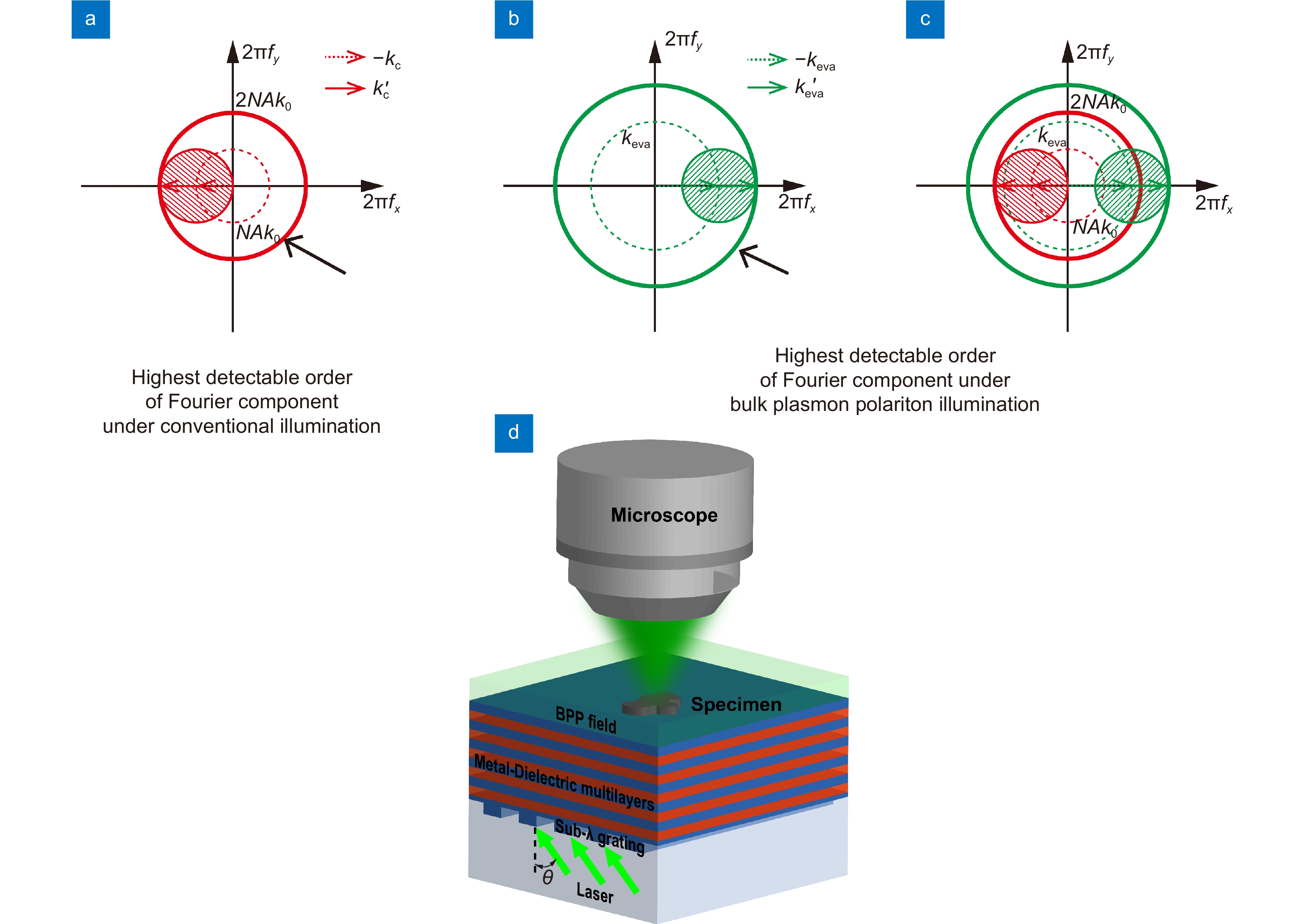
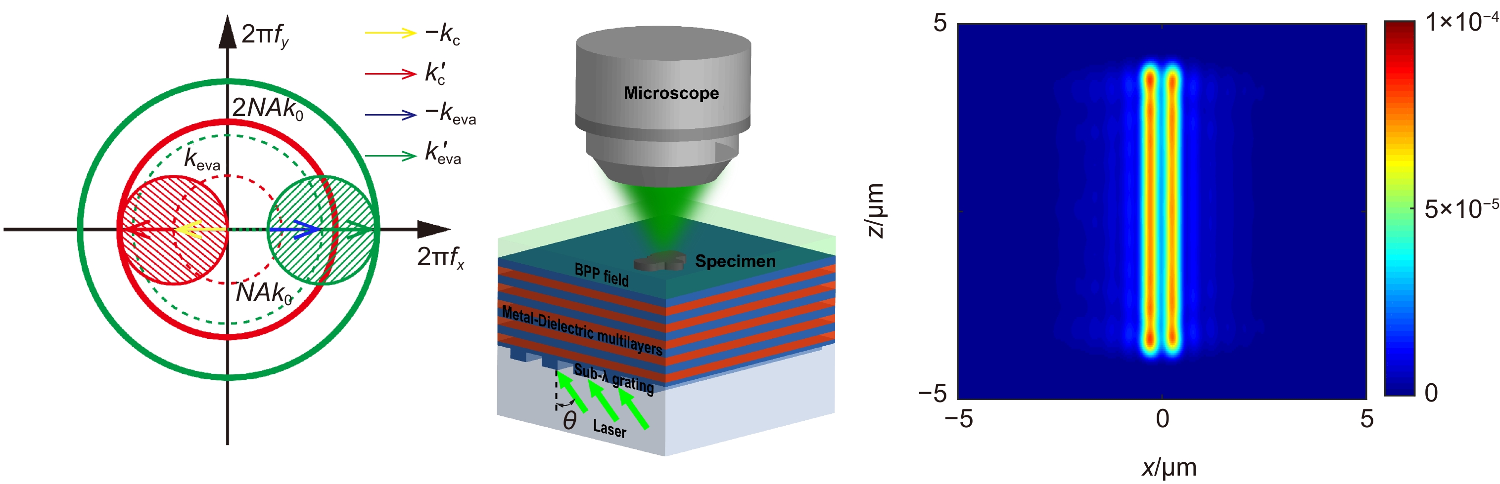
图 1 BPP照明移频超分辨成像示意图。(a) 传统照明下傅里叶分量探测范围示意图,其中kc与k'c分别为传统显微成像中照明光与散射光的横向波矢;(b) BPP照明下傅里叶分量探测范围示意图,其中keva与k'eva分别为BPP照明下照明光与散射光的横向波矢;(c) 最高可探测傅里叶分量的扩展(从红色实线到绿色实线),频谱平移原理示意图;(d) BPP照明显微成像示意图
Figure 1. Schematic diagram of frequency shift super-resolution imaging under BPP illumination. (a) Schematic diagram of fourier component detection range under conventional illumination,kc and k'c are transverse wave vectors of illuminating and scattered light, respectively, under conventional illumination; (b) Schematic diagram of fourier component detection range under BPP illumination, keva and k'eva are transverse wave vectors of illuminating and scattered light, respectively, under BPP illumination; (c) Extension of the highest detectable fourier component (from red solid line to green solid line), schematic diagram of frequency shift effect; (d) Schematic of BPP illumination
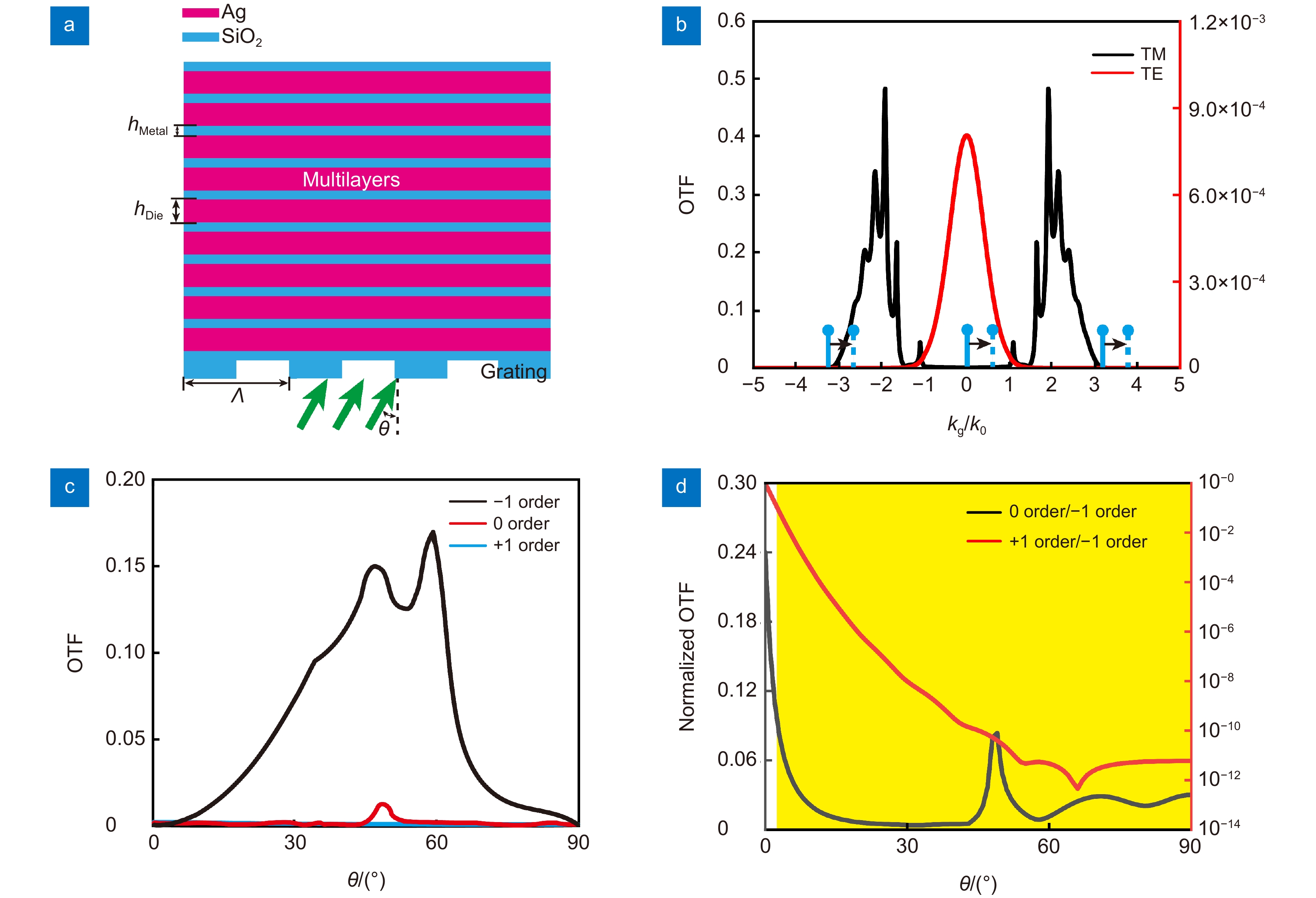
图 2 BPP激发结构。(a) BPP激发结构示意图;(b) TM与TE偏振下Ag/SiO2多层膜OTF;(c) −1、0与+1级次的OTF;(d)不同衍射级次的OTF比值
Figure 2. BPP structure. (a) Schematic of the BPP structure; (b) OTF of Ag/SiO2 multilayers in TM and TE polarization; (c) OTF for −1、0 and +1 orders; (d) The ratio of OTF for different diffraction orders
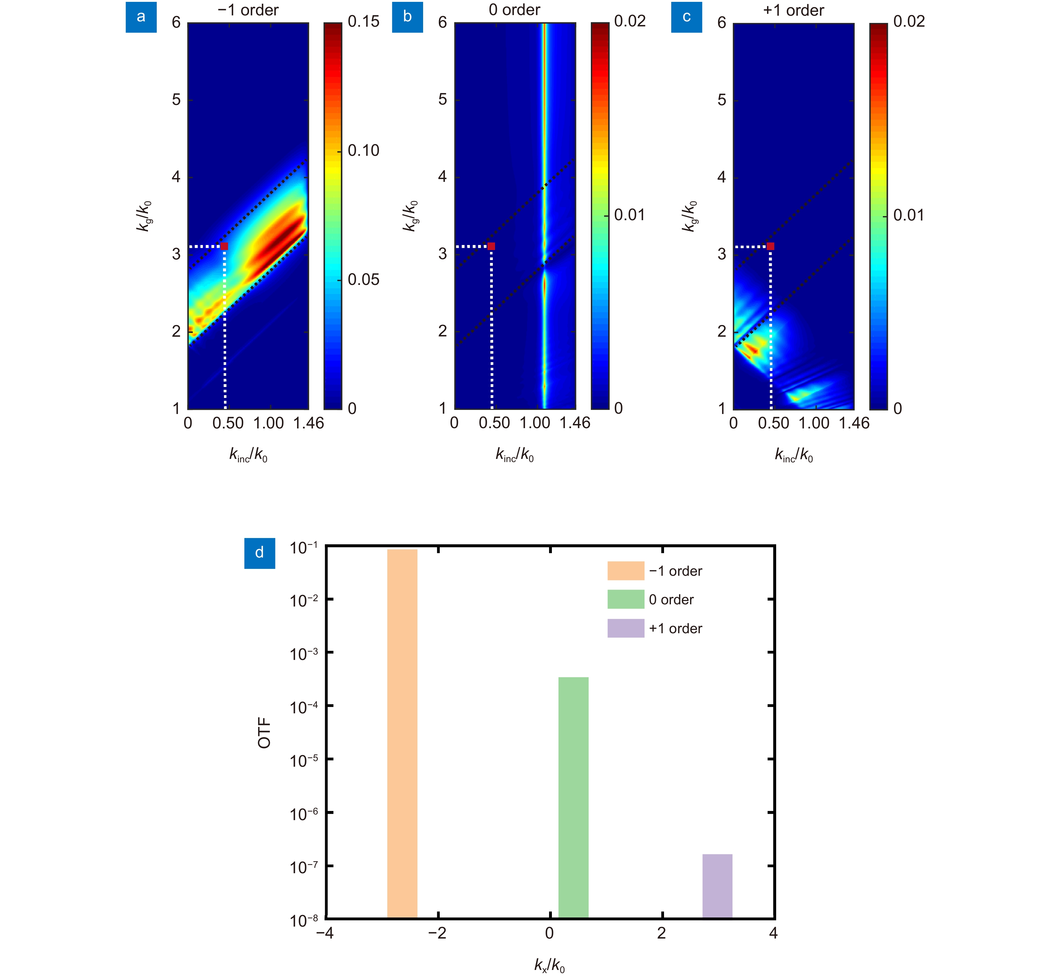
图 3 (a)~(c) 分别为针对不同激发光栅波矢kg和照明光横向波矢kinc,−1级次、0级次以及+1级次的OTF;(d) 光栅周期为170 nm,入射角度为18°,−1、0与+1级次的OTF
Figure 3. (a)~(c) The OTF of −1, 0, and +1 orders for for different kinc and kg, respectively; (d) The OTF of −1, 0, and +1 orders with 170 nm pitch grating at the incident angle of 18 degree
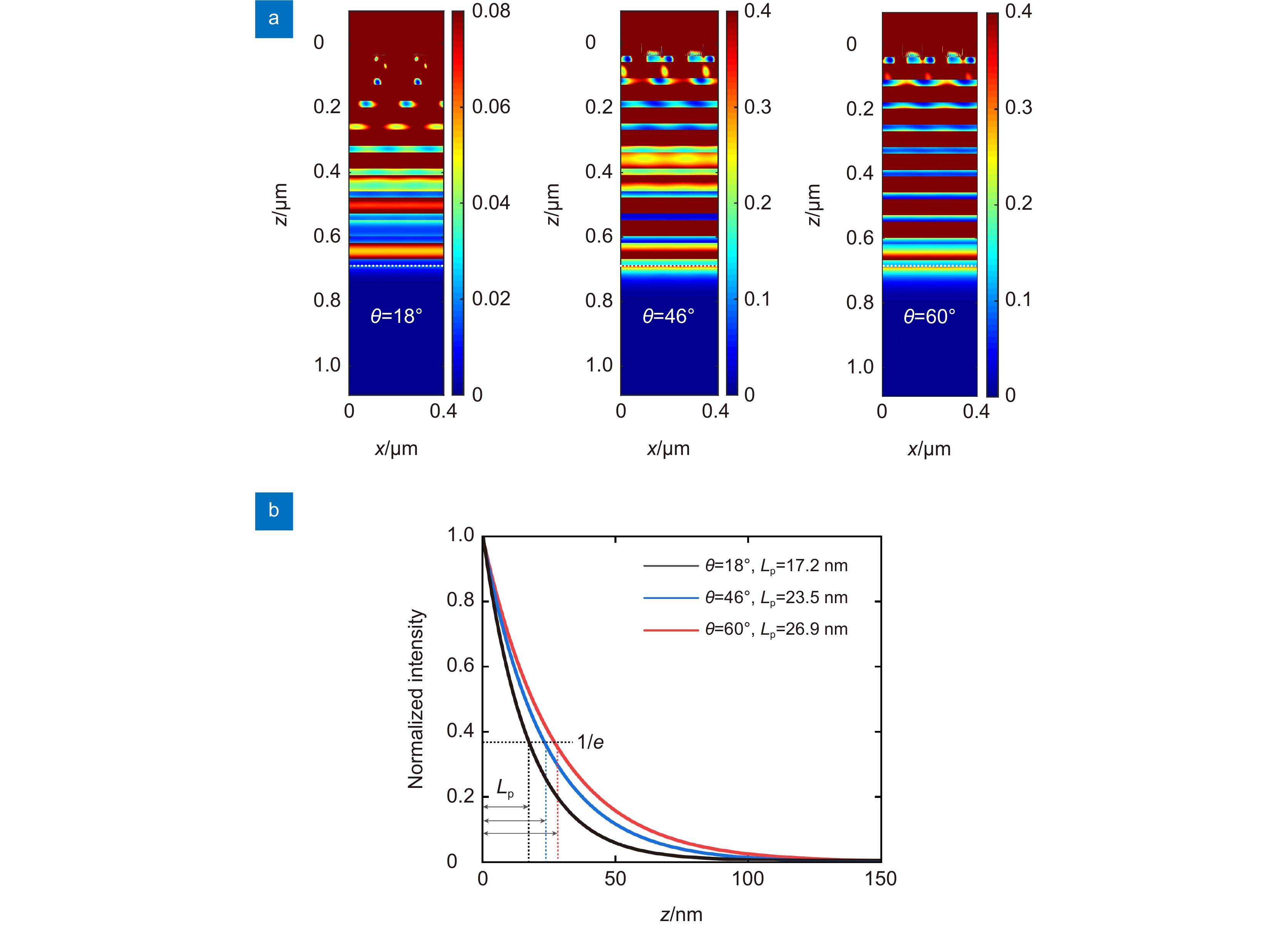
图 4 (a) 入射角为18°、46°和60°时截面光强分布,其中虚线为出射界面;(b) 在18°、46°和60°入射角度下BPP照明光强衰减曲线及穿透深度
Figure 4. (a) The optical intensity distributions in the x–z plane when incidence angles are 18°, 46° and 60°, respectively; (b) The corresponding intensity decay curves away from the illumination surface in (a)
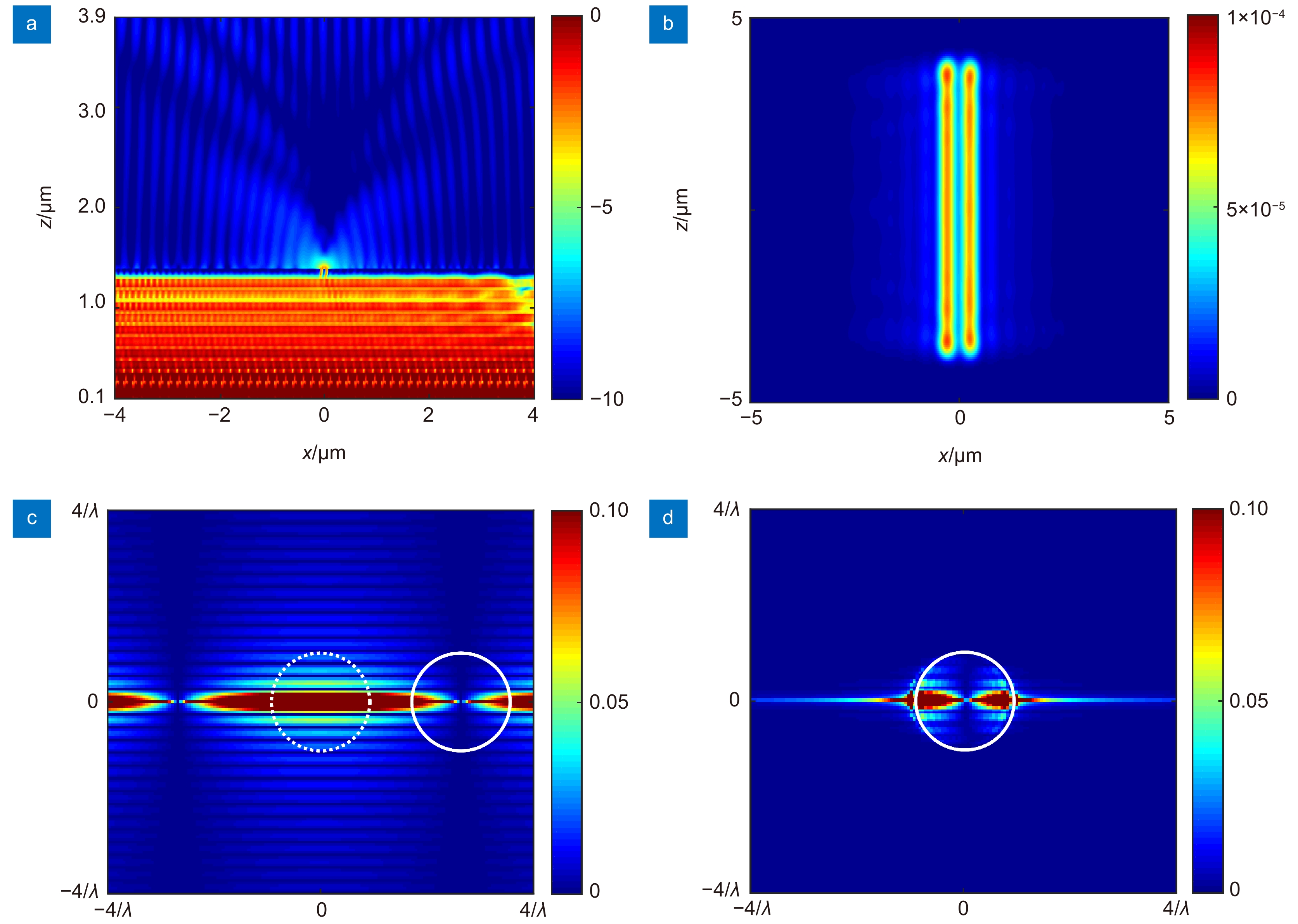
图 5 中心距100 nm双缝结构在2.66k0 BPP照明下的远场成像仿真过程与结果。(a) 采用FDTD方法截面处监视器记录的BPP结构表面倏逝波被散射到远场的x-z截面光强分布;(b) 中心距100 nm双缝结构的远场成像光强分布;(c) 双缝结构的频谱;(d) 远场探测到的双缝结构散射光频谱
Figure 5. Far-field imaging simulation processes and results for double-slit structure with center-to center distance of 100 nm under BPP illumination with 2.66k0. (a) The intensity distribution in the x–z plane in FDTD method; (b) Far-field imaging intensity for double-slit structure; (c) Spatial spectrum of double-slit structures; (d) Spatial spectrum of scattered light for double-slit structures in the far field
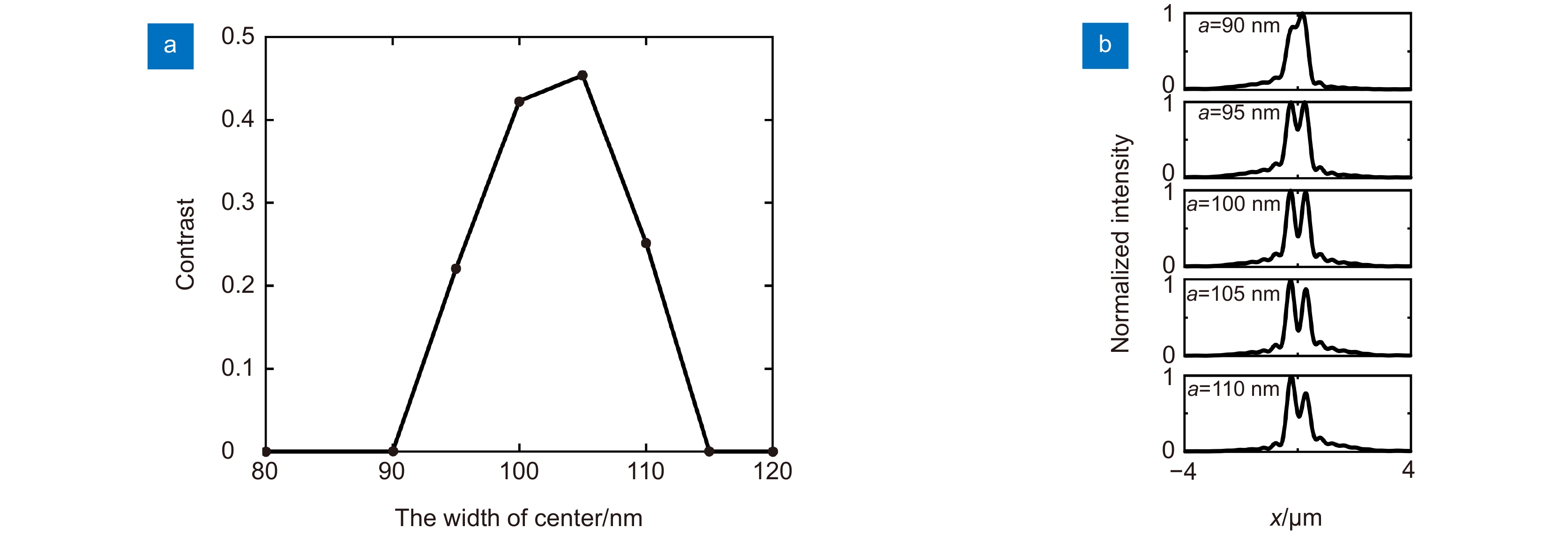
图 6 (a) BPP照明下不同双缝中心距远场成像对比度;(b) 中心距90 nm~110 nm的双缝结构远场成像场强归一化
Figure 6. (a) The contrast of far-field imaging for double-slit structure with different center-to-center distances illuminated by BPP; (b) The normalized optical intensity for double-slit structure with different center-to-center distances in far-field imaging
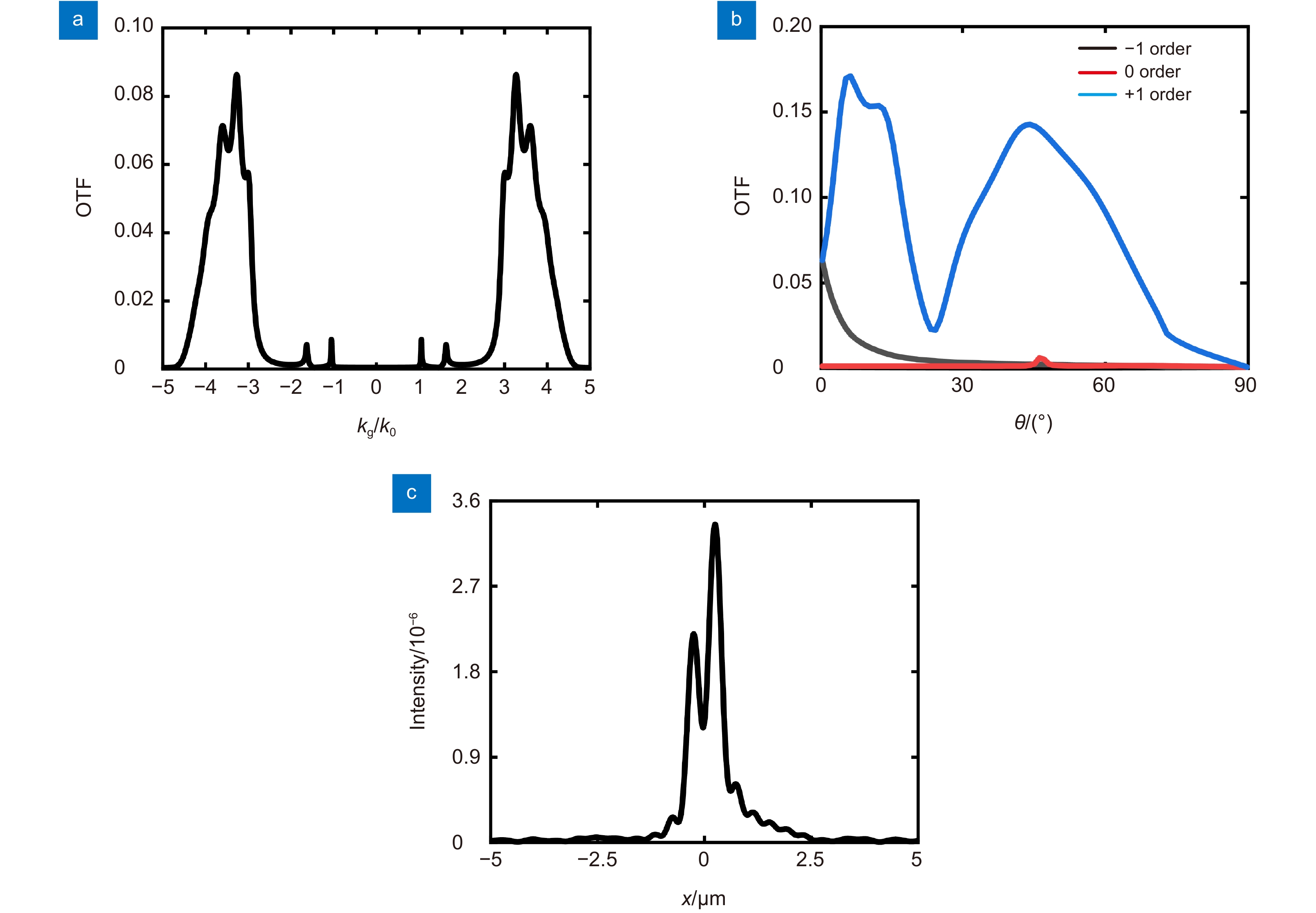
图 7 横向波矢3.86k0的BPP照明超分辨成像。(a) TM偏振入射HMM的OTF;(b) −1、0与+1级次的OTF;(c) 中心距68 nm双缝结构的远场成像光强分布
Figure 7. Super-resolution imaging under BPP illumination with transverse wave vector 3.86k0. (a) OTF of Ag/SiO2 multilayers in TM polarization; (b) OTF for −1、0 and +1 orders; (c) Far-field imaging intensity for double-slit structure
表 1 已被报道基于移频原理的远场无标记超分辨显微成像技术主要参数
Table 1. Main parameters reported based on frequency shift principle label-free far-field subdiffraction imaging techniques
Work Material Resolution Appl.Phys.Lett. 102, 013104 (2013) Micro-fiber ~λ/3.56 (λ=800 nm, NA=0.8) Opt Lett. 38(14), 2455 (2013) TIR Prism ~λ/2.5 (λ=600 nm, NA=0.8) Phys Rev Lett. 118, 076101 (2017) Fluorescent NW ~λ/3.7 (λ=520 nm, NA=0.85) Opt Lett. 46(6), 1265 (2021) Microparticle ~λ/1.68 (λ=505 nm, NA=0.9) BPP illumination HMM ~λ/7.82 (λ=532 nm, NA=0.85)  下载: 导出CSV
-
参考文献
[1] Leung B O, Chou K C. Review of super-resolution fluorescence microscopy for biology[J]. Appl Spectrosc, 2011, 65(9): 967−980. doi: 10.1366/11-06398
[2] Betzig E, Patterson G H, Sougrat R, et al. Imaging intracellular fluorescent proteins at nanometer resolution[J]. Science, 2006, 313(5793): 1642−1645. doi: 10.1126/science.1127344
[3] Hell S W, Wichmann J. Breaking the diffraction resolution limit by stimulated emission: stimulated-emission-depletion fluorescence microscopy[J]. Opt Lett, 1994, 19(11): 780−782. doi: 10.1364/OL.19.000780
[4] Boneberg J, Leiderer P. Optical near-field imaging and nanostructuring by means of laser ablation[J]. Opto-Electron Sci, 2022, 1(1): 210003. doi: 10.29026/oes.2022.210003
[5] Lewis A, Isaacson M, Harootunian A, et al. Development of a 500 Å spatial resolution light microscope: I. light is efficiently transmitted through λ/16 diameter apertures[J]. Ultramicroscopy, 1984, 13(3): 227−231. doi: 10.1016/0304-3991(84)90201-8
[6] 刘小威. 基于照明调控的无标记远场超分辨显微成像[D]. 杭州: 浙江大学, 2017: 7–11.
Liu X W. Label-free far-field super resolution microscopy based on illumination modulation[D]. Hangzhou: Zhejiang University, 2017: 7–11.
[7] Axelrod D, Thompson N L, Burghardt T P. Total internal reflection fluorescent microscopy[J]. J Microsc, 1983, 129(1): 19−28. doi: 10.1111/j.1365-2818.1983.tb04158.x
[8] Farinas J, Simanek V, Verkman A S. Cell volume measured by total internal reflection microfluorimetry: application to water and solute transport in cells transfected with water channel homologs[J]. Biophys J, 1995, 68(4): 1613−1620. doi: 10.1016/S0006-3495(95)80335-8
[9] Cox I J, Sheppard C J R. Scanning optical microscope incorporating a digital framestore and microcomputer[J]. Appl Opt, 1983, 22(10): 1474−1478. doi: 10.1364/AO.22.001474
[10] White J G, Amos W B, Fordham M. An evaluation of confocal versus conventional imaging of biological structures by fluorescence light microscopy[J]. J Cell Biol, 1987, 105(1): 41−48. doi: 10.1083/jcb.105.1.41
[11] Klar T A, Jakobs S, Dyba M, et al. Fluorescence microscopy with diffraction resolution barrier broken by stimulated emission[J]. Proc Natl Acad Sci USA, 2000, 97(15): 8206−8210. doi: 10.1073/pnas.97.15.8206
[12] Hell S W, Kroug M. Ground-state-depletion fluorscence microscopy: a concept for breaking the diffraction resolution limit[J]. Appl Phys B, 1995, 60(5): 495−497. doi: 10.1007/BF01081333
[13] Rust M J, Bates M, Zhuang X W. Sub-diffraction-limit imaging by stochastic optical reconstruction microscopy (STORM)[J]. Nat Methods, 2006, 3(10): 793−796. doi: 10.1038/nmeth929
[14] Betzig E. Proposed method for molecular optical imaging[J]. Opt Lett, 1995, 20(3): 237−239. doi: 10.1364/OL.20.000237
[15] Schermelleh L, Carlton P M, Haase S, et al. Subdiffraction multicolor imaging of the nuclear periphery with 3D structured illumination microscopy[J]. Science, 2008, 320(5881): 1332−1336. doi: 10.1126/science.1156947
[16] Gustafsson M G L. Surpassing the lateral resolution limit by a factor of two using structured illumination microscopy[J]. J Microsc, 2000, 198(2): 82−87. doi: 10.1046/j.1365-2818.2000.00710.x
[17] Wei F F, Liu Z W. Plasmonic structured illumination microscopy[J]. Nano Lett, 2010, 10(7): 2531−2536. doi: 10.1021/nl1011068
[18] Wei F F, Lu D L, Shen H, et al. Wide field super-resolution surface imaging through plasmonic structured illumination microscopy[J]. Nano Lett, 2014, 14(8): 4634−4639. doi: 10.1021/nl501695c
[19] Zheng G A, Horstmeyer R, Yang C. Wide-field, high-resolution fourier ptychographic microscopy[J]. Nat Photonics, 2013, 7(9): 739−745. doi: 10.1038/nphoton.2013.187
[20] Hao X, Liu X, Kuang C F, et al. Far-field super-resolution imaging using near-field illumination by micro-fiber[J]. Appl Phys Lett, 2013, 102(1): 013104. doi: 10.1063/1.4773572
[21] Hao X, Kuang C F, Li Y H, et al. Evanescent-wave-induced frequency shift for optical super resolution imaging[J]. Opt Lett, 2013, 38(14): 2455−2458. doi: 10.1364/OL.38.002455
[22] Liu X W, Kuang C F, Hao X, et al. Fluorescent nanowire ring illumination for wide-field far-field subdiffraction imaging[J]. Phys Rev Lett, 2017, 118(7): 076101. doi: 10.1103/PhysRevLett.118.076101
[23] Ling J Z, Wang Y C, Liu X, et al. Resolution improvement of dark-field microscopy via microparticle near-field illumination[J]. Opt Lett, 2021, 46(6): 1265−1268. doi: 10.1364/OL.418159
[24] Song M W, Wang D, Kudyshev Z A, et al. Enabling optical steganography, data storage, and encryption with plasmonic colors[J]. Laser Photon Rev, 2021, 15(3): 2000343. doi: 10.1002/lpor.202000343
[25] Wang Y L, Fan Q B, Xu T. Design of high efficiency achromatic metalens with large operation bandwidth using bilayer architecture[J]. Opto-Electron Adv, 2021, 4(1): 200008. doi: 10.29026/oea.2021.200008
[26] Zhu Y C, Chen X L, Yuan W Z, et al. A waveguide metasurface based quasi-far-field transverse-electric superlens[J]. Opto-Electron Adv, 2021, 4(10): 210013. doi: 10.29026/oea.2021.210013
[27] Wang H T, Hao C L, Lin H, et al. Generation of super-resolved optical needle and multifocal array using graphene oxide metalenses[J]. Opto-Electron Adv, 2021, 4(2): 200031. doi: 10.29026/oea.2021.200031
[28] 周毅, 梁高峰, 温中泉, 等. 光学超分辨平面超构透镜研究进展[J]. 光电工程, 2021, 48(12): 210399. doi: 10.12086/oee.2021.210399
Zhou Y, Liang G F, Wen Z Q, et al. Recent research progress in optical super-resolution planar meta-lenses[J]. Opto-Electron Eng, 2021, 48(12): 210399. doi: 10.12086/oee.2021.210399
[29] Yue Z, Li J T, Li J, et al. Terahertz metasurface zone plates with arbitrary polarizations to a fixed polarization conversion[J]. Opto-Electron Sci, 2022, 1(3): 210014. doi: 10.29026/oes.2022.210014
[30] Luo X G, Ishihara T. Surface plasmon resonant interference nanolithography technique[J]. Appl Phys Lett, 2004, 84(23): 4780−4782. doi: 10.1063/1.1760221
[31] Luo X G, Ishihara T. Subwavelength photolithography based on surface-plasmon polariton resonance[J]. Opt Express, 2004, 12(14): 3055−3065. doi: 10.1364/OPEX.12.003055
[32] Pu M B, Guo Y H, Li X, et al. Revisitation of extraordinary young’s interference: from catenary optical fields to spin-orbit interaction in metasurfaces[J]. ACS Photonics, 2018, 5(8): 3198−3204. doi: 10.1021/acsphotonics.8b00437
[33] Luo X G, Pu M B, Li X, et al. Young’s double-slit interference enabled by surface plasmon polaritons: a review[J]. J Phys D Appl Phys, 2020, 53(5): 053001. doi: 10.1088/1361-6463/ab50cd
[34] Fang N, Lee H, Sun C, et al. Sub-diffraction-limited optical imaging with a silver superlens[J]. Science, 2005, 308(5721): 534−537. doi: 10.1126/science.1108759
[35] Wang C T, Zhao Z Y, Gao P, et al. Surface plasmon lithography beyond the diffraction limit[J]. Chin Sci Bull, 2016, 61(6): 585−599. doi: 10.1360/N972015-01038
[36] Luo X G. Extraordinary Young’s Interferences and Super-Diffraction Laser Lithography[M]//Sugioka K. Handbook of Laser Micro- and Nano-Engineering. Cham: Springer, 2020: 1–40.
[37] Gao P, Yao N, Wang C T, et al. Enhancing aspect profile of half-pitch 32 nm and 22 nm lithography with plasmonic cavity lens[J]. Appl Phys Lett, 2015, 106(9): 093110. doi: 10.1063/1.4914000
[38] Beliaev L Y, Takayama O, Melentiev P N, et al. Photoluminescence control by hyperbolic metamaterials and metasurfaces: a review[J]. Opto-Electron Adv, 2021, 4(8): 210031. doi: 10.29026/oea.2021.210031
[39] Schurig D, Smith D R. Spatial filtering using media with indefinite permittivity and permeability tensors[J]. Appl Phys Lett, 2003, 82(14): 2215−2217. doi: 10.1063/1.1562344
[40] Wood B, Pendry J B, Tsai D P. Directed subwavelength imaging using a layered metal-dielectric system[J]. Phys Rev B, 2006, 74(11): 115116. doi: 10.1103/PhysRevB.74.115116
[41] Wang C T, Gao P, Tao X, et al. Far field observation and theoretical analyses of light directional imaging in metamaterial with stacked metal-dielectric films[J]. Appl Phys Lett, 2013, 103(3): 031911. doi: 10.1063/1.4815924
[42] Guo Z, Zhao Z Y, Yan L S, et al. Moiré fringes characterization of surface plasmon transmission and filtering in multi metal-dielectric films[J]. Appl Phys Lett, 2014, 105(14): 141107. doi: 10.1063/1.4896022
[43] Liu Z W, Lee H, Xiong Y, et al. Far-field optical hyperlens magnifying sub-diffraction-limited objects[J]. Science, 2007, 315(5819): 1686. doi: 10.1126/science.1137368
[44] Liu H C, Kong W J, Liu K P, et al. Deep subwavelength interference lithography with tunable pattern period based on bulk plasmon polaritons[J]. Opt Express, 2017, 25(17): 20511−20521. doi: 10.1364/OE.25.020511
[45] Kong W J, Du W J, Liu K P, et al. Launching deep subwavelength bulk plasmon polaritons through hyperbolic metamaterials for surface imaging with a tuneable ultra-short illumination depth[J]. Nanoscale, 2016, 8(38): 17030−17038. doi: 10.1039/C6NR03313J
[46] Kong W, Du W J, Liu K P, et al. Surface imaging microscopy with tunable penetration depth as short as 20 nm by employing hyperbolic metamaterials[J]. J Mater Chem C, 2018, 6(7): 1797−1805. doi: 10.1039/C7TC04748G
[47] 杜文娟, 王长涛, 赵泽宇, 等. 金属-介质多层膜超衍射材料频谱滤波特性[J]. 光电工程, 2014, 41(11): 89−94. doi: 10.3969/j.issn.1003-501X.2014.11.015
Du W J, Wang C T, Zhao Z Y, et al. Characteristic investigation of spatial spectrums filtering in metal-dielectric multilayer metamaterials[J]. Opto-Electron Eng, 2014, 41(11): 89−94. doi: 10.3969/j.issn.1003-501X.2014.11.015
[48] Born M, Wolf E. Principles of Optics: Electromagnetic Theory of Propagation, Interference and Diffraction of Light[M]. 7th ed. Cambridge: Cambridge University, 1999.
[49] Palik E D. Handbook of Optical Constants of Solids. Volume III[M]. San Diego: Academic Press, 1998: 356, 760.
-
访问统计
 点击扫一扫
点击扫一扫
图(8)
表(1)
计量
- 文章访问数:
- PDF下载数:
- 施引文献: 0


