
 E-mail Alert
 RSS
RSS
-
摘要
内窥镜图像质量在医生对早期病灶、异型增生复发的诊断中至关重要。因此本文根据血管对光谱的吸收特性,提出了一种基于光谱变换的血管增强算法。首先,该算法对图像RGB通道进行导向滤波,将各通道分为亮度层和细节层;接着,将各通道的细节层进行基于信噪比的自适应增强,并将亮度层进行拉伸,使得GB通道的信息增强,R通道信息降低;最后,将各通道合并生成增强图像。本文应用该算法对大量内窥镜图像进行增强,并且与Spectra B增强技术相比较。本文方法在DV-BV指标和韦伯对比度指标均优于Spectra B。

Abstract
Endoscopic image quality plays an important role in the diagnosis of early lesions and dysplasia. Therefore, a blood vessel enhancement algorithm based on spectral absorption characteristics of blood vessels is proposed in this paper. First of all, RGB channels are obtained from the color image and divided into the brightness layer with the high dynamic range and the detail layer with the detail image information through the guided filter. Then, the detail layer of each channel is adaptively enhanced based on SNR (signal noise ratio), and the brightness layer is stretched to improve the GB channel information and to reduce R channel information. Finally each channel is merged to generate an enhanced image. In this article, a large number of endoscopic images is applied to this algorithm and compared with Karl Stroz's Spectra B enhancement technology. This method performs better in image enhancements while using the Detail Variance-Background Variance index and the Weber contrast index to evaluate.
-
Key words:
- endoscope /
- guided filter /
- vessel enhancement /
- spectral enhancement
-
Overview

Overview: With the development of the minimally invasive surgery, endoscopes have become the necessary medical devices that permit the endoscopists to examine the gastrointestinal mucosa and identify the abnormal tissue. However, early diseases are overlooked and tumors still remain in the conventional white light endoscopic surgery. Foreign companies have put many special image enhancement algorithms forward, while there is a lack of this function in domestic products. In order to solve the above problems, the special image enhancement algorithms are very important for endoscopes.
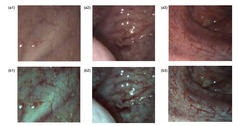
This paper proposes a blood vessel enhancement algorithm based on the optical spectral absorption characteristics of blood vessels. The contrasts of capillaries and vessels are highlighted by means of reducing the red spectral reflection and increasing the blue and green spectral reflection. The enhancement algorithm includes two aspects: the detail enhancement and the brightness enhancement. Firstly, RGB channels are obtained from the color image and divided into the brightness layer with the high dynamic range and the detail layer with the detail image information through the guided filter. Then, each pixel of the detail image multiplies by an enhanced factor, and the factor of each channel is calculated based on SNR (signal noise ratio). The improvement factor can improve the quality of image enhancement, but excessive factor will amplify the image noise. To get the stretched factor using in brightness layer, each channel is converted from RGB space to CIE space. In this paper, the distance is calculated between the blood vessel and the background in a series of the representative oral vascular biomedical images taken by endoscope (including before and after the image enhancement), and the stretching coefficient is obtained after averaging. After that, the brightness layer is stretched to enhance the GB channel information and to reduce R channel information. Blood vessel information is highlighted because the color of background region turns to green and white, while the color of vessels turns to red and dark. Finally, the detail enhanced image and the brightness-enhanced image are merged to generate a enhanced image.
In order to evaluate the validity of the proposed enhancement method, this paper uses the detail variance-background variance (DV-BV) index and Weber contrast index. For evaluating the enhancement algorithm, the algorithm has been applied to a large number of images captured by endoscopes. The assessment of subjective and objective indicators shows the significant enhancements. Moreover, compared with Karl Stroz's Spectra B enhancement technology, the method proposed in this paper performs better in image enhancement.
-

-
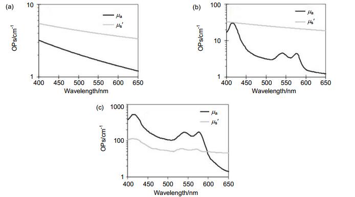
图 1 吸收系数(μa)和散射系数(μs′)。
Figure 1. Absorption coefficient (μa) and scattering coefficient (μs′).
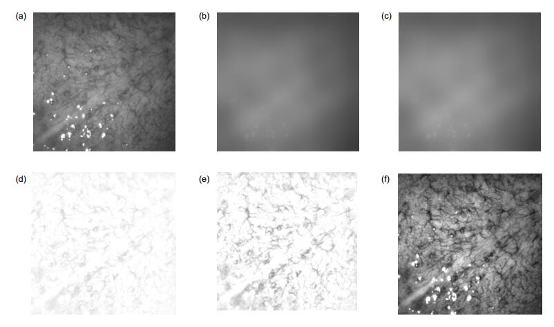
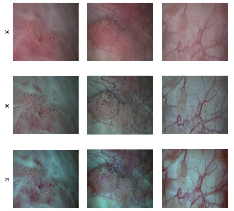
图 3 G 通道对比度增强算法效果图。
Figure 3. Endoscopic images of G channel after contrast enhancement.
表 1 DV-BV指标与韦伯对比度指标
Table 1. DV-BV index and Weber contrast index
Image No. DV-BV index Weber contrast index Original Enhanced Original Enhanced 1 10.11 16.75 0.08 0.18 2 13.15 15.85 0.18 0.29 3 11.13 13.94 0.12 0.2  下载: 导出CSV
表 2 DV-BV指标与韦伯对比度指标和Spectra B对比
Table 2. DV-BV index and Weber contrast index compared with Spectra B
Image No. DV-BV index Weber contrast index Original Karl Storz Our method Original Karl Storz Our method 1 5.34 9.42 10.91 0.1 0.12 0.12 2 4.69 7.85 9.39 0.16 0.2 0.23 3 5.25 8.61 10.62 0.2 0.23 0.24
下载: 导出CSV
-
参考文献
[1] Li B P, Meng Max Q H. Wireless capsule endoscopy images enhancement via adaptive contrast diffusion[J]. Journal of Visual Communication and Image Representation, 2012, 23(1): 222-228. doi: 10.1016/j.jvcir.2011.10.002
[2] Gono K. Narrow band imaging: technology basis and research and development history[J]. Clinical Endoscopy, 2015, 48(6): 476-480. doi: 10.5946/ce.2015.48.6.476
[3] Togashi K, Osawa H, Koinuma K, et al. A comparison of conventional endoscopy, chromoendoscopy, and the optimal-band imaging system for the differentiation of neoplastic and non-neoplastic colonic polyps[J]. Gastrointestinal Endoscopy, 2009, 69(3): 734-741. doi: 10.1016/j.gie.2008.10.063
[4] Kamphuis G M, de Bruin D M, Fallert T, et al. Storz professional image enhancement system: a new technique to improve endoscopic bladder imaging[J]. Journal of Cancer Science & Therapy, 2016, 8(3): 71-77.
[5] Okuhata H, Nakamura H, Hara S, et al. Application of the real-time Retinex image enhancement for endoscopic images[C]//Proceedings of the 2013 35th Annual International Conference of the IEEE Engineering in Medicine and Biology Society, 2013: 3407-3410.
[6] Gopi V P, Palanisamy P. Capsule endoscopic image denoising based on double density dual tree complex wavelet transform[J]. International Journal of Imaging and Robotics, 2013, 9(1): 48-60. http://en.cnki.com.cn/Article_en/CJFDTotal-GXJM200905037.htm
[7] Imtiaz M S, Mohammed S K, Deeba F, et al. Tri-Scan: A three stage color enhancement tool for endoscopic images[J]. Journal of Medical Systems, 2017, 41(6): 102. doi: 10.1007/s10916-017-0738-z
[8] Du Le V N, Wang Q Z, Gould T, et al. Vascular contrast in narrow-band and white light imaging[J]. Applied Optics, 2014, 53(18): 4061-4071. doi: 10.1364/AO.53.004061
[9] Noorden G K V. The Theory of Binocular Vision[J]. American Journal of Ophthalmology, 1977, 84(5):751-751. 10.1016/0002-9394(77)90403-2
[10] 许轰烈, 陈钱, 顾国华, 等.利用导向滤波的宽动态范围图像增强技术[J].红外与激光工程, 2015, 44(12): 3843-3849. doi: 10.3969/j.issn.1007-2276.2015.12.058
Xu H L, Chen Q, Gu G H, et al. High dynamic range image enhancement technology based on guided image filter[J]. Infrared and Laser Engineering, 2015, 44(12): 3843-3849. doi: 10.3969/j.issn.1007-2276.2015.12.058
[11] Ghimire D, Lee J. Nonlinear transfer function-based local approach for color image enhancement[J]. IEEE Transactions on Consumer Electronics, 2011, 57(2): 858-865. doi: 10.1109/TCE.2011.5955233
[12] Gono K, Obi T, Yamaguchi M, et al. Appearance of enhanced tissue features in narrow-band endoscopic imaging[J]. Journal of Biomedical Optics, 2004, 9(3): 568-577. doi: 10.1117/1.1695563
[13] Otsu N. A threshold selection method from gray-level histograms[J]. Automatica, 1975, 11(285-296): 23-27. http://ieeexplore.ieee.org/xpls/abs_all.jsp?arnumber=4310076
-
访问统计
 点击扫一扫
点击扫一扫
图(7)
表(2)
计量
- 文章访问数:
- PDF下载数:
- 施引文献: 0


