
 E-mail Alert
 RSS
RSS
-
摘要:
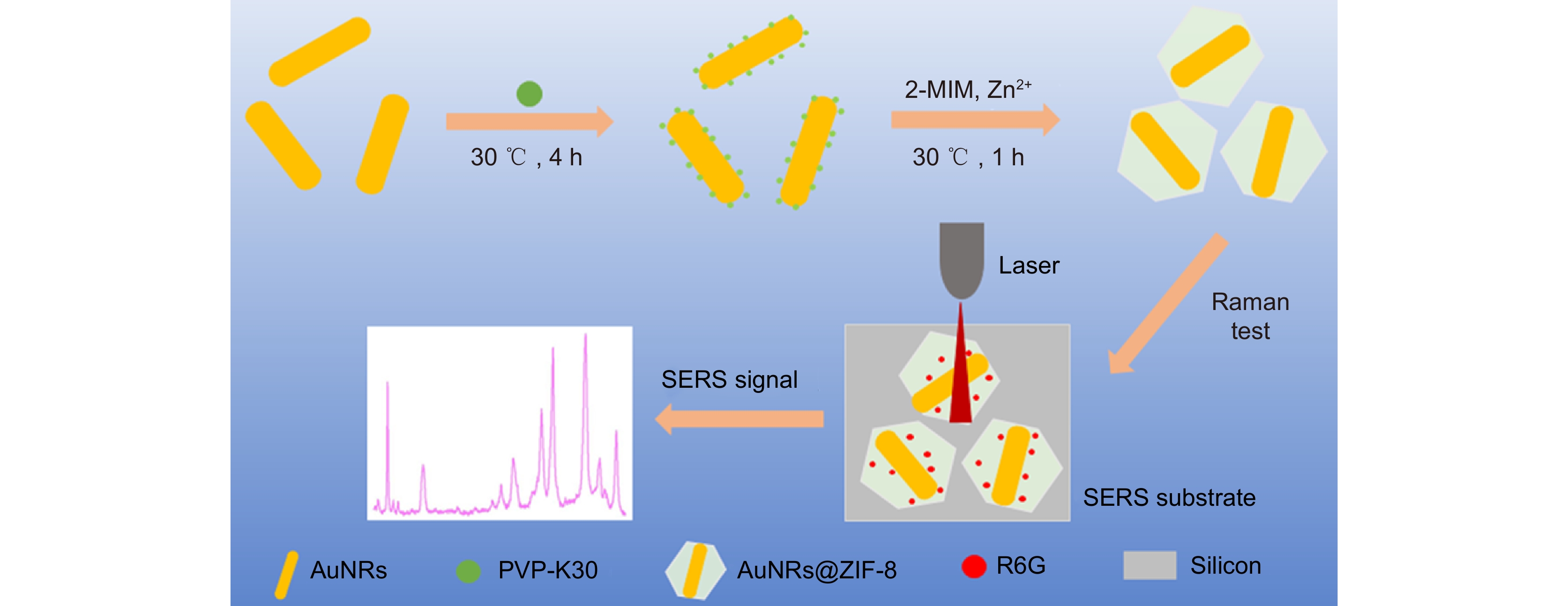
表面增强拉曼光谱(Surface enhanced Raman spectroscopy, SERS)是一种分子光谱,具有快速、高灵敏和指纹识别的特性,在分析化学、生物医学等领域有着重要的应用。然而,在溶液样品中一些检测分子很难被SERS基底所吸附,导致分子拉曼信号增强困难。为此,本文提出了一种ZIF-8材料包覆金纳米棒(AuNRs)的核壳结构(AuNRs@ZIF-8)来实现拉曼信号增强,既可利用金纳米颗粒的表面等离激元增强特性,又可利用ZIF-8这种多孔MOFs材料的吸附性能,从而实现溶液样品的高灵敏拉曼检测。我们首先采用晶种法制备了均一性良好的AuNRs,然后对其进行聚乙烯吡咯烷酮(PVP)修饰,最后加入金属有机框架ZIF-8前驱体,得到AuNRs@ZIF-8核壳纳米结构。该结构对罗丹明(R6G)的SERS检测灵敏度很高,检测限可低至10−9 mol/L,并且线性关系和均一性均良好。此外,我们通过测试该结构吸收R6G前后的UV-Vis吸收光谱进一步证实了核壳纳米结构的生成和对目标分子的有效吸附。
-
关键词:
- SERS /
- AuNRs@ZIF-8 /
- 核壳纳米结构 /
- 光吸收 /
- 吸附性
Abstract:Surface enhanced Raman spectroscopy (SERS) is a kind of molecular spectrum, which has the characteristics of rapidity, high sensitivity and fingerprint recognition, and has important applications in analytical chemistry, biomedicine and other fields. However, some detection molecules in the solution sample are difficult to be adsorbed by the SERS substrate, resulting in difficulty in enhancing the Raman signal of molecular. To this end, this paper proposes a core-shell structure (AuNRs@ZIF-8) of ZIF-8 material coated gold nanorods (AuNRs) to achieve Raman signal enhancement. Both the surface plasmon enhancement characteristics of the gold nanoparticles and the adsorption properties of ZIF-8, a porous MOFs material, can be used to realize highly sensitive Raman detection of the solution samples. We first prepare the well-homogeneous AuNRs by seed crystallization method, then modify them with polyvinylpyrrolidone (PVP), and finally add the metal-organic framework ZIF-8 precursor to obtain the AuNRs@ZIF-8 Core-shell nanostructures. The structure has high sensitivity to the SERS detection of rhodamine (R6G), the detection limit can be as low as 10−9 M, and the linear relationship and homogeneity are good. In addition, we further confirm the formation of the core-shell nanostructures and effective adsorption of the target molecules by testing the UV-Vis absorption spectra of the structure before and after R6G absorption.
-
Key words:
- SERS /
- AuNRs@ZIF-8 /
- Core-shell nanostructure /
- light absorption /
- adsorption
-
Overview: As a non-destructive, rapid and highly sensitive molecular detection technology, surface enhanced Raman spectroscopy (SERS) has been widely used in analytical chemistry, biomedicine, food detection and other fields. Currently, it is generally believed that there are two kinds of SERS enhanced mechanism. One is the chemical mechanism of charge transfer between molecules. The second is the physical mechanism, namely local surface plasmon resonance (LSPR). When the incident laser is resonantly coupled with the metal substrate (Au, Ag, Cu, etc.) at a certain excitation wavelength, a strong local electromagnetic field is formed, which is confined to a small range on the metal surface. This phenomenon, known as local surface plasmon resonance (LSPR), can greatly enhance the Raman signals of the detected molecules. Generally, only when the detected molecules are close to the surface of the precious metal structure through chemical or physical action could there be a good SERS signal. Therefore, it has become particularly important to design SERS bases with strong adsorption capacity for detected molecules.
Metal-organic framework (MOFs) is a new type of porous framework material which is self-assembled by metal ions or clusters as nodes and organic molecules as ligands through coordination bonds. Due to its unique network structure, large porosity, flexible structure, and multiple molecular adsorption sites, MOFs are very conducive to sample enrichment, and are widely used in adsorption, separation, and storage of solution samples. Combining metal organic framework with metal substrate material, using MOFs enrichment and SERS effect of precious metals, the detection molecules could be close to the surface of precious metals, thus effectively collecting Raman signals of the test molecules.
This paper proposes a core-shell structure (AuNRs@ZIF-8) of ZIF-8 material coated gold nanorods (AuNRs) to achieve Raman signal enhancement. Both the surface plasmon enhancement characteristics of the gold nanoparticles and the adsorption properties of ZIF-8, a porous MOFs material, can be used to realize highly sensitive Raman detection of the solution samples. We first prepare well-homogeneous AuNRs by seed crystallization method, then modify them with polyvinylpyrrolidone (PVP), and finally add the metal-organic framework ZIF-8 precursor to obtain the AuNRs@ZIF-8 Core-shell nanostructures. The structure has high sensitivity to the SERS detection of rhodamine (R6G), the detection limit can be as low as 10−9 M, and the linear relationship and homogeneity are good. In addition, we further confirm the formation of the core-shell nanostructures and effective adsorption of the target molecules by testing the UV-Vis absorption spectra of the structure before and after R6G absorption.
-

-
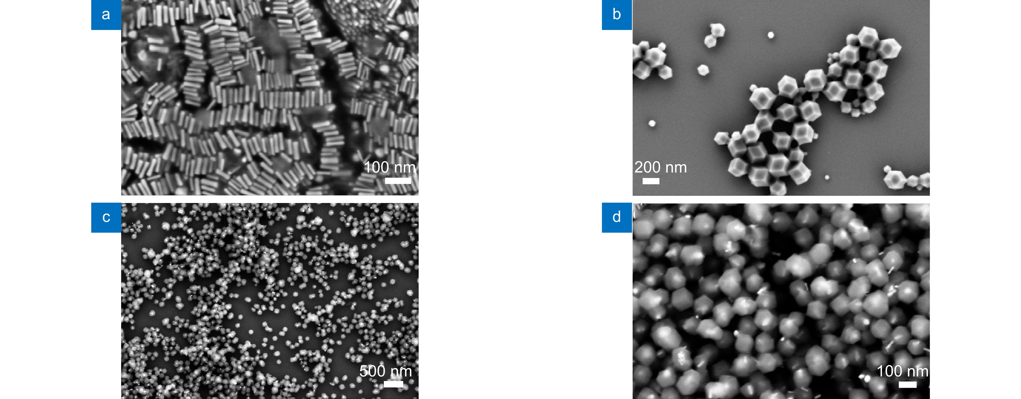
图 2 样品SEM图片。(a)金纳米棒;(b) ZIF-8;(c, d) 不同倍率下AuNRs@ZIF-8核壳纳米粒子
Figure 2. SEM images of (a) AuNRs, (b) ZIF -8, (c, d) AuNRs@ZIF-8 of different magnifications
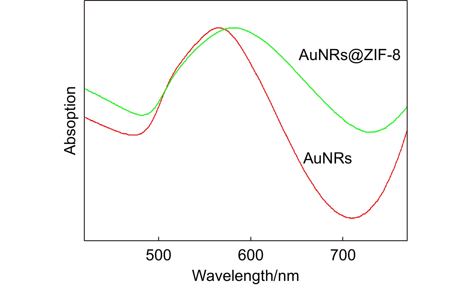
图 3 AuNRs@ZIF-8和AuNRs的UV-Vis光谱图
Figure 3. UV–Vis absorption spectra of the as-synthesized AuNRs and AuNRs@ZIF-8
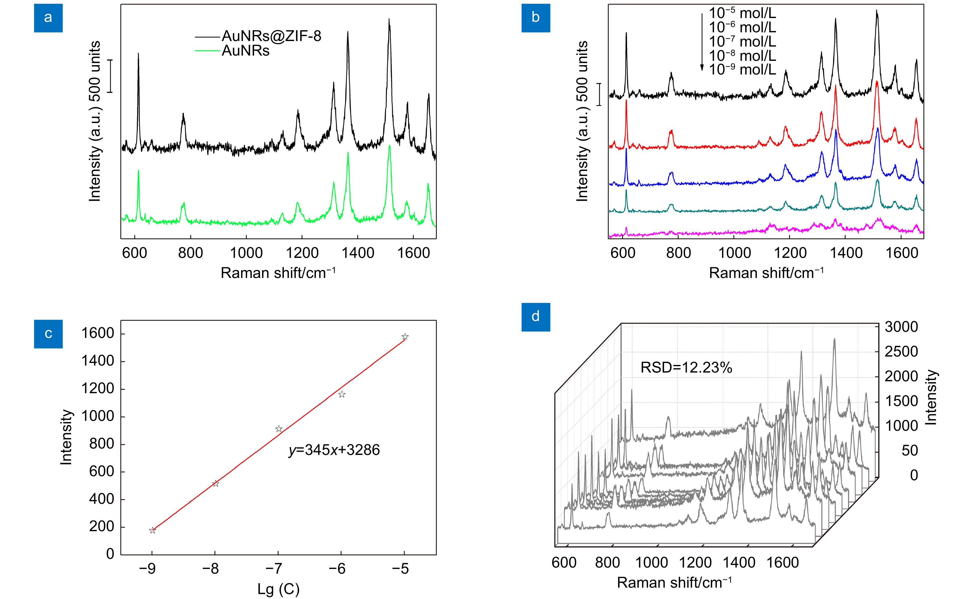
图 4 样品SERS性能图。(a) AuNRs@ZIF-8和AuNRs对10−5 mol/L的R6G的SERS光谱;(b) AuNRs@ZIF-8 SERS基底对10−9至10−4 mol/L浓度范围的R6G的SERS光谱;(c) R6G的621 cm−1处的拉曼强度与不同R6G浓度的对数值的关系拟合;(d) AuNRs@ZIF-8 SERS基底对浓度为10−4 mol/L的R6G十个不同位置的SERS光谱图
Figure 4. Sample performance diagram. (a) SERS spectra of 10−5 mol/L R6G for AuNRs@ZIF-8 and AuNRs substrates; (b) SERS spectra of R6G with the concentration ranging from 10−9 to 10−4 mol/L for AuNRs@ZIF-8 substrates; (c) The Raman intensities at 612 cm−1 versus logarithmic values of R6G concentrations; (d) Series of SERS spectra of random 10 sites of the substrates absorbing 10−4 mol/L R6G
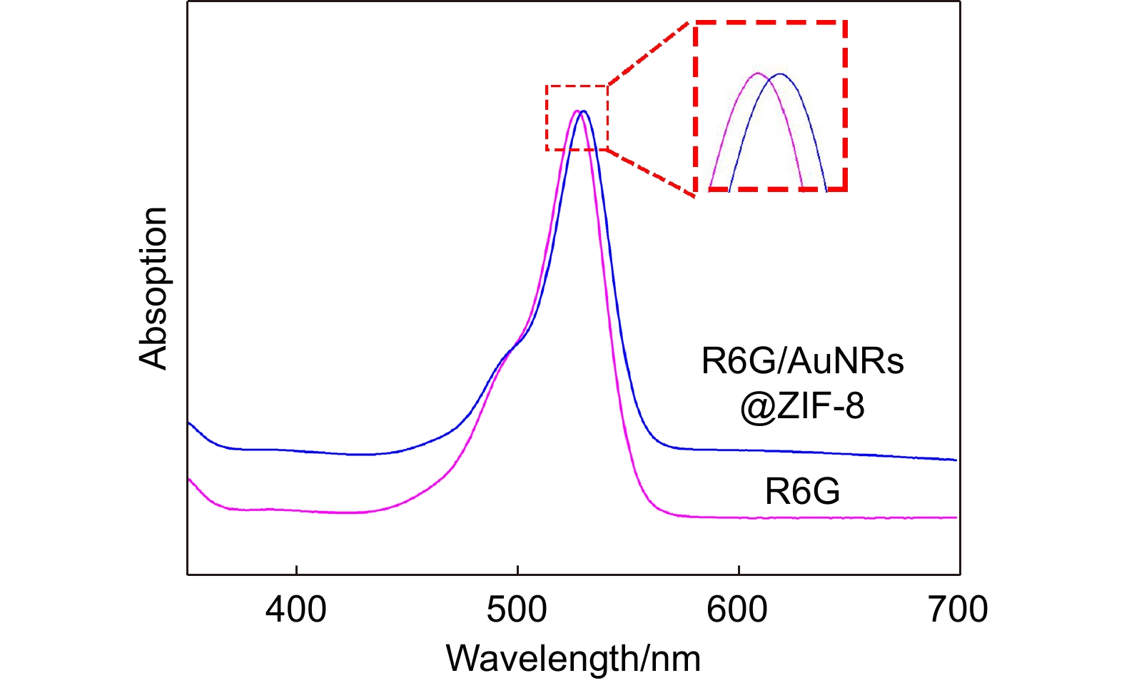
图 6 R6G溶液和R6G/AuNRs@ZIF-8混合溶液的UV-Vis光谱图
Figure 6. UV–vis absorption spectra of the R6G and R6G/AuNRs@ZIF-8
-
[1] Luo X G. Engineering Optics 2.0: A Revolution in Optical Theories, Materials, Devices and Systems[M]. Singapore: Springer, 2019. https://doi.org/10.1007/978-981-13-5755-8.
[2] Bharati M S S, Soma V R. Flexible SERS substrates for hazardous materials detection: recent advances[J]. Opto-Electron Adv, 2021, 4(11): 210048. doi: 10.29026/oea.2021.210048
[3] Nie B B, Luo Y Y, Shi J P, et al. Bowl-like Pore array made of hollow Au/Ag alloy nanoparticles for SERS detection of melamine in solid milk powder[J]. Sens Actuators B Chem, 2019, 301: 127087. doi: 10.1016/j.snb.2019.127087
[4] Luo X G, Tsai D, Gu M, et al. Extraordinary optical fields in nanostructures: from sub-diffraction-limited optics to sensing and energy conversion[J]. Chem Soc Rev, 2019, 48(8): 2458−2494. doi: 10.1039/C8CS00864G
[5] 王向贤, 陈函文, 朱剑凯, 等. 金纳米锥阵列与金薄膜耦合结构表面等离子体折射率传感研究[J]. 光电工程, 2022, 49(12): 220135. doi: 10.12086/oee.2022.220135
Wang X X, Chen H W, Zhu J K, et al. Research on surface plasmon refractive index sensing of gold nano cone array and gold film coupling structure[J]. Opto-Electron Eng, 2022, 49(12): 220135. doi: 10.12086/oee.2022.220135
[6] 魏欣然, 梁瑜章, 何怡瑾, 等. 传感品质因数增强的塔姆-表面等离激元杂化模式[J]. 光电工程, 2022, 49(11): 220217. doi: 10.12086/oee.2022.220217
Wei X R, Liang Y Z, He Y J, et al. Tamm-surface plasmon hybrid mode for improving sensing figure of merit[J]. Opto-Electron Eng, 2022, 49(11): 220217. doi: 10.12086/oee.2022.220217
[7] Du X, Xiong L, Zhao X Q, et al. Dual-band bound states in the continuum based on hybridization of surface lattice resonances[J]. Nanophotonics, 2022, 11(21): 4843−4853. doi: 10.1515/nanoph-2022-0427
[8] Huang C H, Li A L, Chen X Y, et al. Understanding the role of metal-organic frameworks in surface-enhanced raman scattering application[J]. Small, 2020, 16(43): 2004802. doi: 10.1002/smll.202004802
[9] Osterrieth J W M, Wright D, Noh H, et al. Core-shell gold nanorod@zirconium-based metal-organic framework composites as in situ size-selective raman probes[J]. J Am Chem Soc, 2019, 141(9): 3893−3900. doi: 10.1021/jacs.8b11300
[10] He J C, Dong J W, Hu Y F, et al. Design of Raman tag-bridged core-shell Au@Cu3(BTC)2 nanoparticles for Raman imaging and synergistic chemo-photothermal therapy[J]. Nanoscale, 2019, 11(13): 6089−6100. doi: 10.1039/C9NR00041K
[11] Zhang X L, Li G L, Wu D, et al. Recent progress in the design fabrication of metal-organic frameworks-based nanozymes and their applications to sensing and cancer therapy[J]. Biosens Bioelectron, 2019, 137: 178−198. doi: 10.1016/j.bios.2019.04.061
[12] Chen Q Q, Hou R N, Zhu Y Z, et al. Au@ZIF-8 core-shell nanoparticles as a SERS substrate for volatile organic compound gas detection[J]. Anal Chem, 2021, 93(19): 7188−7195. doi: 10.1021/acs.analchem.0c05432
[13] Li Q Q, Gong S S, Zhang H, et al. Tailored necklace-like Ag@ZIF-8 core/shell heterostructure nanowires for high-performance plasmonic SERS detection[J]. Chem Eng J, 2019, 371: 26−33. doi: 10.1016/j.cej.2019.03.236
[14] Zheng G C, De Marchi S, López-Puente V, et al. Encapsulation of single plasmonic nanoparticles within ZIF-8 and SERS analysis of the MOF flexibility[J]. Small, 2016, 12(29): 3935−3943. doi: 10.1002/smll.201600947
[15] De Marchi S, Vázquez-Iglesias L, Bodelón G, et al. Programmable modular assembly of functional proteins on raman-encoded zeolitic imidazolate framework-8 (ZIF-8) nanoparticles as SERS tags[J]. Chem Mater, 2020, 32(13): 5739−5749. doi: 10.1021/acs.chemmater.0c01518
[16] Huang J F, Xu Z R, Jiang Y H, et al. Metal organic framework-coated gold nanorod as an on-demand drug delivery platform for chemo-photothermal cancer therapy[J]. J Nanobiotechnology, 2021, 19(1): 219. doi: 10.1186/s12951-021-00961-x
[17] Li R, Li W, Jin C, et al. Fabrication of ZIF-8@TiO2 micron composite via hydrothermal method with enhanced absorption and photocatalytic activities in tetracycline degradation[J]. J Alloys Compd, 2020, 825: 154008. doi: 10.1016/j.jallcom.2020.154008
[18] Weng C C, Luo Y Y, Wang B F, et al. Layer-dependent SERS enhancement of TiS2 prepared by simple electrochemical intercalation[J]. J Mater Chem C, 2020, 8(40): 14138−14145. doi: 10.1039/D0TC03683H
[19] Yang Y, Ma X X, Yang C H, et al. Eco-friendly and acid-resistant magnetic porous carbon derived from ZIF-67 and corn stalk waste for effective removal of imidacloprid and thiamethoxam from water[J]. Chem Eng J, 2022, 430: 132999. doi: 10.1016/J.CEJ.2021.132999
[20] Yuan Y T, Gao L, Luo Y Y, et al. Ag@ZIF-67 nanocomposites for ultra-sensitive SERS detection to thiram molecules[J]. J Phys D Appl Phys, 2023, 56(5): 055302. doi: 10.1088/1361-6463/acadea
-
 下载:
下载:


 点击扫一扫
点击扫一扫
图(7)
计量
- 文章访问数:
- PDF下载数:
- 施引文献: 0


