
 E-mail Alert
 RSS
RSS
-
摘要:
传统落射式荧光显微镜的探测光路和照明光路处于同轴位置,成像质量会受到非焦平面荧光的影响。光片荧光显微镜(Light sheet fluorescence microscopy,LSFM)区别于传统的荧光显微镜,它的探测光路和照明光路呈直角排布,照明光为一个薄片,成像时只有光片区域的样本被照亮,这种照明方式能够有效降低非焦平面荧光激发。同时,激光每次只照亮一个平面,能够有效降低样本的照射时间,由此降低光毒性和光漂白性的影响。本文首先介绍了光片荧光显微镜的基本光路组成结构,以及在这些结构基础上进行的优化创新;之后介绍了针对离体样本和活体样本发展出的多种解决方案。得益于这些创新,光片荧光显微镜能够在较长时间范围内对荧光标记的生物样本进行3D成像。最后提出了光片荧光显微镜发展的潜在方向以及局限性,希望能给研究人员提供更为系统的光片荧光显微镜方面的知识以及一些有益的参考。
Abstract:In a traditional epi-illumination fluorescence microscope, the detection path is coaxial with the illumination path, which induces the non-focal plane fluorescence and deteriorates the imaging quality. Light sheet fluorescence microscopy (LSFM), differing from the traditional fluorescence microscopes, adopts an orthogonal configuration of detection and illumination paths. A thin sheet is formed from the excitation beam, which only excites a single layer of the sample. This methodology prevents the excited fluorescence from the non-focal plane during imaging. Besides, the utilization of the laminar illumination light can significantly reduce the exposure time of the fluorescence imaging. As a result, the effects of photobleaching and phototoxicity are decreased. In this review, we first introduce the basic light path structure compositions of a LSFM system as well as the optimization and innovation based on these structures. Next, we discuss enormous processing methods developed for samples both in vitro and in vivo. Benefiting from all these innovations, LSFM outstands in performing the 3D imaging of the fluorescence-labeled biological samples and can function steadily for a long recording time. Finally, we propose potential researching directions in the future, and discuss the technical limitations of current LSFM. This review aims to provide researchers in the relevant scientific research fields with a comprehensive understanding and inspiring reference of LSFM techniques.
-
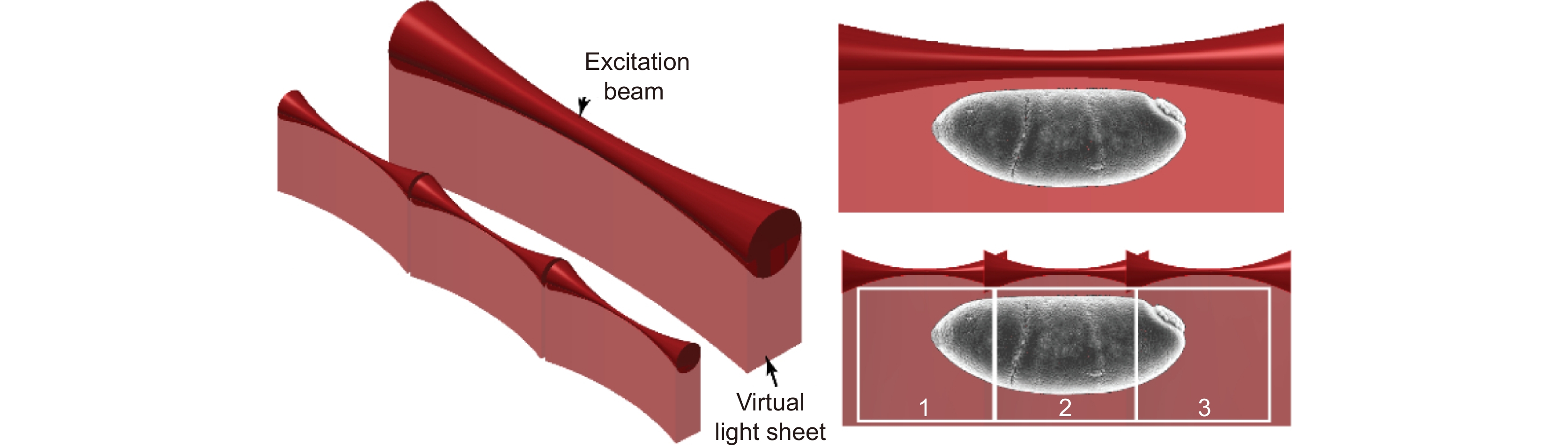
Overview: Light sheet fluorescence microscopy (LSFM), as a type of fluorescence microscope, can image fluorescence-labelled specific ribosomes, proteins, and cells, and observe their positions and functions in biological tissues. Different from traditional fluorescence microscopes, LSFM’s detection path and illumination path are arranged at orthogonal orientation. The excitation beam is a thin sheet, only a slice region of the sample is illuminated, thus reducing the fluorescence generation in the non-focal plane. Besides only a single plane is illuminated by the laser at a time, which significantly reduces the exposure time of the fluorescent molecules, thereby minimizing the effects of photobleaching and phototoxicity. Due to these properties, LSFM can perform 3D imaging of fluorescence-labeled biological samples for a long recording time. Nowadays, it has been widely used in many biological fields such as neuroscience, developmental biology, and histopathology.
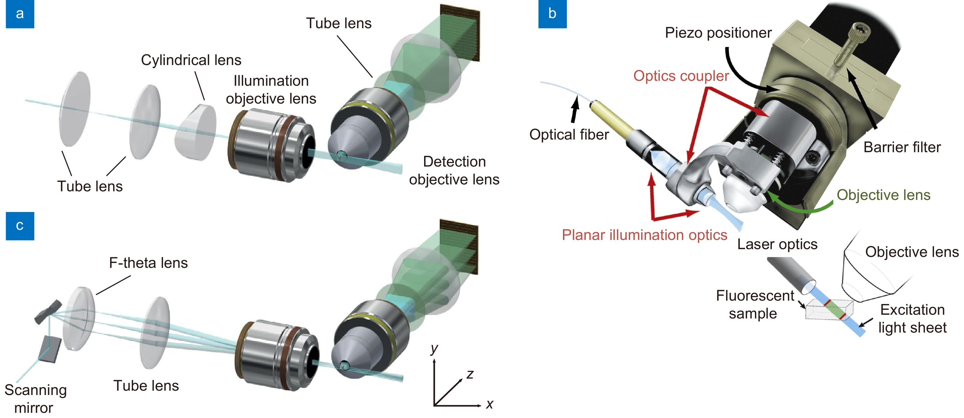
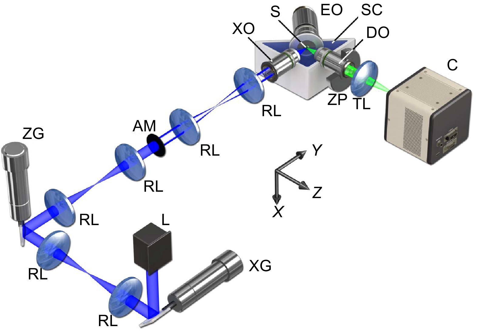
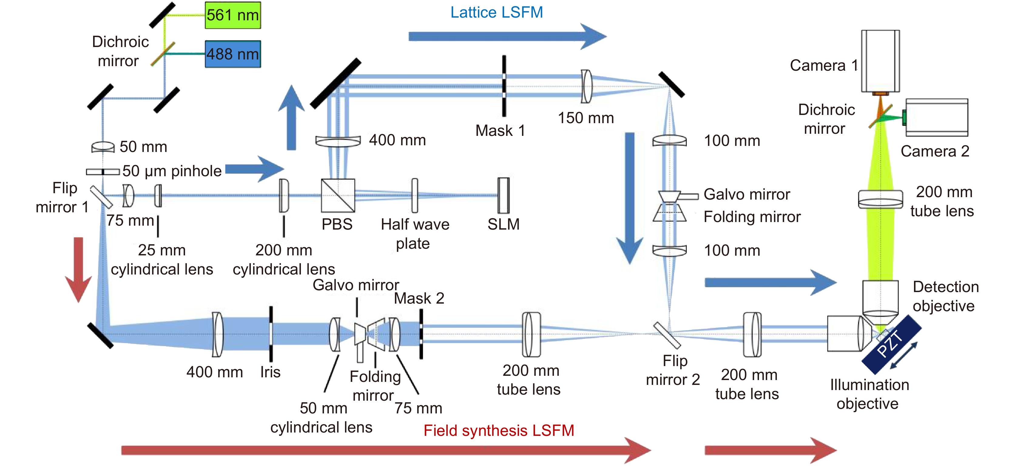
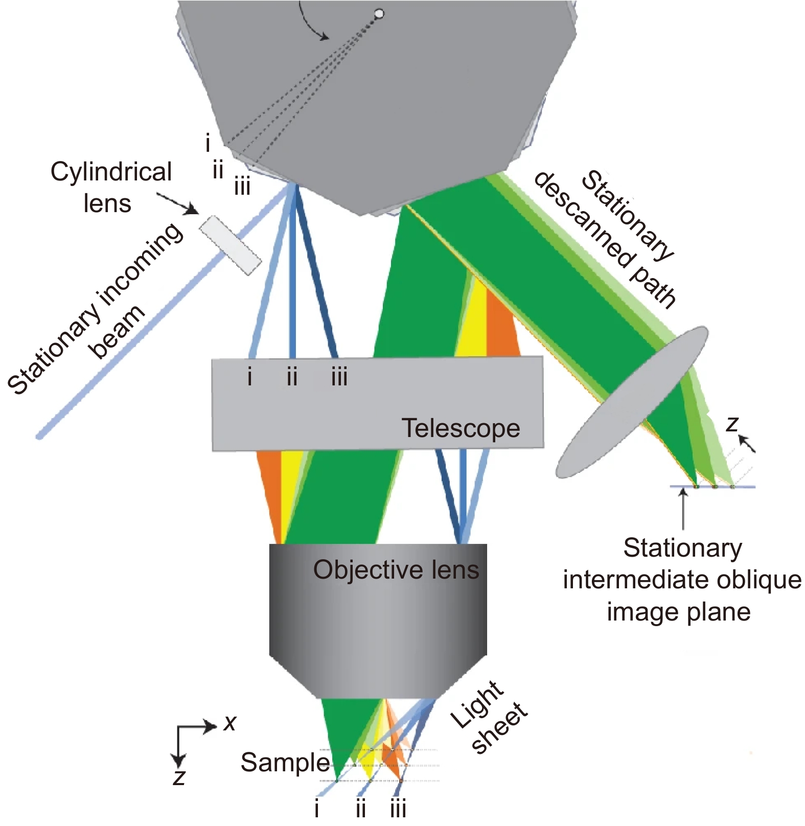
In the first part, the LSFM with classical optical path configurations, such as SPIM, OCPI, and DSLM are described from the perspective of optical path construction. The work made by researchers to promote the resolution and imaging throughput based on those work are introduced. These methods include changing the beam structure, shortening the optical path distance, and increasing the imaging speed, many of which are still beneficial to us today. In the second part, fluorescent dyes and immunofluorescence staining techniques for biological samples used in LSFM are described, including tissue transparency and sample fixing of living animals. These sample processing methods have greatly promoted the development of fluorescence microscopes, and representative studies are listed.
Finally, the review summarizes both the advantages and disadvantages of LSFM as well as the potential development direction and limitations. The orthogonal optical path configuration limits the lateral size of the sample, and the imaging performance is poor for the opaque or high-scattering samples. LSFM has higher requirements for both the size and transparency of the sample. It is considered that the breakthrough of the LSFM in future breakthroughs mainly lies in two aspects: improving the imaging parameters and adapting to more biological applications. It should be done from a biological point of view, in conjunction with other technologies, to advance the development of LSFM. Finally, this review is expected to provide researchers with a more systematic knowledge of light-sheet fluorescence microscopy and some useful references.
-

-
-
[1] Stephens D J, Allan V J. Light microscopy techniques for live cell imaging[J]. Science, 2003, 300(5616): 82−86. doi: 10.1126/science.1082160
[2] Truong T V, Supatto W, Koos D S, et al. Deep and fast live imaging with two-photon scanned light-sheet microscopy[J]. Nat Methods, 2011, 8(9): 757−760. doi: 10.1038/nmeth.1652
[3] Siedentopf H, Zsigmondy R. Uber sichtbarmachung und größenbestimmung ultramikoskopischer teilchen, mit besonderer anwendung auf goldrubingläser[J]. Ann Phys, 1902, 315(1): 1−39. doi: 10.1002/andp.19023150102
[4] Voie A H, Burns D H, Spelman F A. Orthogonal-plane fluorescence optical sectioning: three-dimensional imaging of macroscopic biological specimens[J]. J Microsc, 1993, 170(3): 229−236. doi: 10.1111/j.1365-2818.1993.tb03346.x
[5] Stelzer E H K, Strobl F, Chang B J, et al. Light sheet fluorescence microscopy[J]. Nat Rev Methods Primers, 2021, 1(1): 73. doi: 10.1038/s43586-021-00069-4
[6] Huisken J, Swoger J, Del Bene F, et al. Optical sectioning deep inside live embryos by selective plane illumination microscopy[J]. Science, 2004, 305(5686): 1007−1009. doi: 10.1126/science.1100035
[7] Keller P J, Schmidt A D, Wittbrodt J, et al. Reconstruction of zebrafish early embryonic development by scanned light sheet microscopy[J]. Science, 2008, 322(5904): 1065−1069. doi: 10.1126/science.1162493
[8] Vettenburg T, Dalgarno H I C, Nylk J, et al. Light-sheet microscopy using an Airy beam[J]. Nat Methods, 2014, 11(5): 541−544. doi: 10.1038/nmeth.2922
[9] Planchon T A, Gao L, Milkie D E, et al. Rapid three-dimensional isotropic imaging of living cells using Bessel beam plane illumination[J]. Nat Methods, 2011, 8(5): 417−423. doi: 10.1038/nmeth.1586
[10] Chen B C, Legant W R, Wang K, et al. Lattice light-sheet microscopy: imaging molecules to embryos at high spatiotemporal resolution[J]. Science, 2014, 346(6208): 1257998. doi: 10.1126/science.1257998
[11] Holekamp T F, Turaga D, Holy T E. Fast three-dimensional fluorescence imaging of activity in neural populations by objective-coupled planar illumination microscopy[J]. Neuron, 2008, 57(5): 661−672. doi: 10.1016/j.neuron.2008.01.011
[12] Gao L, Shao L, Chen B C, et al. 3D live fluorescence imaging of cellular dynamics using Bessel beam plane illumination microscopy[J]. Nat Protoc, 2014, 9(5): 1083−1101. doi: 10.1038/nprot.2014.087
[13] Chang B J, Kittisopikul M, Dean K M, et al. Universal light-sheet generation with field synthesis[J]. Nat Methods, 2019, 16(3): 235−238. doi: 10.1038/s41592-019-0327-9
[14] Fiolka R, Stemmer A, Belyaev Y. Virtual slit scanning microscopy[J]. Histochem Cell Biol, 2007, 128(6): 499−505. doi: 10.1007/s00418-007-0342-2
[15] Hosny N A, Seyforth J A, Spickermann G, et al. Planar Airy beam light-sheet for two-photon microscopy[J]. Biomed Opt Express, 2020, 11(7): 3927−3935. doi: 10.1364/BOE.395547
[16] Huisken J, Stainier D Y R. Even fluorescence excitation by multidirectional selective plane illumination microscopy (mSPIM)[J]. Opt Lett, 2007, 32(17): 2608−2610. doi: 10.1364/OL.32.002608
[17] Krzic U, Gunther S, Saunders T E, et al. Multiview light-sheet microscope for rapid in toto imaging[J]. Nat Methods, 2012, 9(7): 730−733. doi: 10.1038/nmeth.2064
[18] Tomer R, Khairy K, Amat F, et al. Quantitative high-speed imaging of entire developing embryos with simultaneous multiview light-sheet microscopy[J]. Nat Methods, 2012, 9(7): 755−763. doi: 10.1038/nmeth.2062
[19] Fu Q Y, Martin B L, Matus D Q, et al. Imaging multicellular specimens with real-time optimized tiling light-sheet selective plane illumination microscopy[J]. Nat Commun, 2016, 7(1): 11088. doi: 10.1038/ncomms11088
[20] Gao L. Extend the field of view of selective plan illumination microscopy by tiling the excitation light sheet[J]. Opt Express, 2015, 23(5): 6102−6111. doi: 10.1364/OE.23.006102
[21] Wang D Y, Jin Y X, Feng R L, et al. Tiling light sheet selective plane illumination microscopy using discontinuous light sheets[J]. Opt Express, 2019, 27(23): 34472−34483. doi: 10.1364/OE.27.034472
[22] Zong W J, Zhao J, Chen X Y, et al. Large-field high-resolution two-photon digital scanned light-sheet microscopy[J]. Cell Res, 2015, 25(2): 254−257. doi: 10.1038/cr.2014.124
[23] Dean K M, Roudot P, Welf E S, et al. Deconvolution-free subcellular imaging with axially swept light sheet microscopy[J]. Biophys J, 2015, 108(12): 2807−2815. doi: 10.1016/j.bpj.2015.05.013
[24] Bouchard M B, Voleti V, Mendes C S, et al. Swept confocally-aligned planar excitation (SCAPE) microscopy for high-speed volumetric imaging of behaving organisms[J]. Nat Photonics, 2015, 9(2): 113−119. doi: 10.1038/nphoton.2014.323
[25] Strnad P, Gunther S, Reichmann J, et al. Inverted light-sheet microscope for imaging mouse pre-implantation development[J]. Nat Methods, 2016, 13(2): 139−142. doi: 10.1038/nmeth.3690
[26] Greer C J, Holy T E. Fast objective coupled planar illumination microscopy[J]. Nat Commun, 2019, 10(1): 4483. doi: 10.1038/s41467-019-12340-0
[27] Fan J T, Suo J L, Wu J M, et al. Video-rate imaging of biological dynamics at centimetre scale and micrometre resolution[J]. Nat Photonics, 2019, 13(11): 809−816. doi: 10.1038/s41566-019-0474-7
[28] Tsien R Y. The green fluorescent protein[J]. Annu Rev Biochem, 1998, 67: 509−544. doi: 10.1146/annurev.biochem.67.1.509
[29] Keppler A, Gendreizig S, Gronemeyer T, et al. A general method for the covalent labeling of fusion proteins with small molecules in vivo[J]. Nat Biotechnol, 2003, 21(1): 86−89. doi: 10.1038/nbt765
[30] Los G V, Encell L P, McDougall M G, et al. HaloTag: a novel protein labeling technology for cell imaging and protein analysis[J]. ACS Chem Biol, 2008, 3(6): 373−382. doi: 10.1021/cb800025k
[31] Shaner N C, Steinbach P A, Tsien R Y. A guide to choosing fluorescent proteins[J]. Nat Methods, 2005, 2(12): 905−909. doi: 10.1038/nmeth819
[32] Matz M V, Fradkov A F, Labas Y A, et al. Fluorescent proteins from nonbioluminescent Anthozoa species[J]. Nat Biotechnol, 1999, 17(10): 969−973. doi: 10.1038/13657
[33] Ai H W, Baird M A, Shen Y, et al. Engineering and characterizing monomeric fluorescent proteins for live-cell imaging applications[J]. Nat Protoc, 2014, 9(4): 910−928. doi: 10.1038/nprot.2014.054
[34] Chen T W, Wardill T J, Sun Y, et al. Ultrasensitive fluorescent proteins for imaging neuronal activity[J]. Nature, 2013, 499(7458): 295−300. doi: 10.1038/nature12354
[35] Kim S Y, Cho J H, Murray E, et al. Stochastic electrotransport selectively enhances the transport of highly electromobile molecules[J]. Proc Natl Acad Sci USA, 2015, 112(46): E6274−E6283. doi: 10.1073/pnas.1510133112
[36] Costantini I, Cicchi R, Silvestri L, et al. In-vivo and ex-vivo optical clearing methods for biological tissues: review[J]. Biomed Opt Express, 2019, 10(10): 5251−5267. doi: 10.1364/BOE.10.005251
[37] Richardson D S, Lichtman J W. Clarifying tissue clearing[J]. Cell, 2015, 162(2): 246−257. doi: 10.1016/j.cell.2015.06.067
[38] Chakraborty T, Driscoll M K, Jeffery E, et al. Light-sheet microscopy of cleared tissues with isotropic, subcellular resolution[J]. Nat Methods, 2019, 16(11): 1109−1113. doi: 10.1038/s41592-019-0615-4
[39] Glaser A K, Reder N P, Chen Y, et al. Multi-immersion open-top light-sheet microscope for high-throughput imaging of cleared tissues[J]. Nat Commun, 2019, 10(1): 2781. doi: 10.1038/s41467-019-10534-0
[40] Dodt H U, Leischner U, Schierloh A, et al. Ultramicroscopy: three-dimensional visualization of neuronal networks in the whole mouse brain[J]. Nat Methods, 2007, 4(4): 331−336. doi: 10.1038/nmeth1036
[41] Hama H, Kurokawa H, Kawano H, et al. Scale: a chemical approach for fluorescence imaging and reconstruction of transparent mouse brain[J]. Nat Neurosci, 2011, 14(11): 1481−1488. doi: 10.1038/nn.2928
[42] Ertürk A, Becker K, Jährling N, et al. Three-dimensional imaging of solvent-cleared organs using 3DISCO[J]. Nat Protoc, 2012, 7(11): 1983−1995. doi: 10.1038/nprot.2012.119
[43] Chung K, Wallace J, Kim S Y, et al. Structural and molecular interrogation of intact biological systems[J]. Nature, 2013, 497(7449): 332−337. doi: 10.1038/nature12107
[44] Park Y G, Sohn C H, Chen R, et al. Protection of tissue physicochemical properties using polyfunctional crosslinkers[J]. Nat Biotechnol, 2019, 37(1): 73−83. doi: 10.1038/nbt.4281
[45] Pan C C, Cai R Y, Quacquarelli F P, et al. Shrinkage-mediated imaging of entire organs and organisms using uDISCO[J]. Nat Methods, 2016, 13(10): 859−867. doi: 10.1038/nmeth.3964
[46] Chen F, Tillberg P W, Boyden E S. Expansion microscopy[J]. Science, 2015, 347(6221): 543−548. doi: 10.1126/science.1260088
[47] Chen F, Wassie A T, Cote A J, et al. Nanoscale imaging of RNA with expansion microscopy[J]. Nat Methods, 2016, 13(8): 679−684. doi: 10.1038/nmeth.3899
[48] Alon S, Goodwin D R, Sinha A, et al. Expansion sequencing: spatially precise in situ transcriptomics in intact biological systems[J]. Science, 2021, 371(6528): eaax2656. doi: 10.1126/science.aax2656
[49] Kaufmann A, Mickoleit M, Weber M, et al. Multilayer mounting enables long-term imaging of zebrafish development in a light sheet microscope[J]. Development, 2012, 139(17): 3242−3247. doi: 10.1242/dev.082586
[50] De Medeiros G, Balázs B, Hufnagel L. Light-sheet imaging of mammalian development[J]. Semin Cell Dev Biol, 2016, 55: 148−155. doi: 10.1016/j.semcdb.2015.11.001
[51] Ichikawa T, Nakazato K, Keller P J, et al. Live imaging of whole mouse embryos during gastrulation: migration analyses of epiblast and mesodermal cells[J]. PLoS One, 2013, 8(7): e64506. doi: 10.1371/journal.pone.0064506
[52] Udan R S, Piazza V G, Hsu C W, et al. Quantitative imaging of cell dynamics in mouse embryos using light-sheet microscopy[J]. Development, 2014, 141(22): 4406−4414. doi: 10.1242/dev.111021
[53] McDole K, Guignard L, Amat F, et al. In toto imaging and reconstruction of post-implantation mouse development at the single-cell level[J]. Cell, 2018, 175(3): 859−876.e33. doi: 10.1016/j.cell.2018.09.031
[54] Hillman E M C, Voleti V, Li W Z, et al. Light-sheet microscopy in neuroscience[J]. Annu Rev Neurosci, 2019, 42: 295−313. doi: 10.1146/annurev-neuro-070918-050357
[55] Wan Y N, McDole K, Keller P J. Light-sheet microscopy and its potential for understanding developmental processes[J]. Annu Rev Cell Dev Biol, 2019, 35: 655−681. doi: 10.1146/annurev-cellbio-100818-125311
[56] Poola P K, Afzal M I, Yoo Y, et al. Light sheet microscopy for histopathology applications[J]. Biomed Eng Lett, 2019, 9(3): 279−291. doi: 10.1007/s13534-019-00122-y
[57] Chang B J, Meza V D P, Stelzer E H K. csiLSFM combines light-sheet fluorescence microscopy and coherent structured illumination for a lateral resolution below 100 nm[J]. Proc Natl Acad Sci USA, 2017, 114(19): 4869−4874. doi: 10.1073/pnas.1609278114
[58] Betzig E, Patterson G H, Sougrat R, et al. Imaging intracellular fluorescent proteins at nanometer resolution[J]. Science, 2006, 313(5793): 1642−1645. doi: 10.1126/science.1127344
[59] Hell S W, Wichmann J. Breaking the diffraction resolution limit by stimulated emission: stimulated-emission-depletion fluorescence microscopy[J]. Opt Lett, 1994, 19(11): 780−782. doi: 10.1364/OL.19.000780
[60] Nelsen E, Hobson C M, Kern M E, et al. Combined atomic force microscope and volumetric light sheet system for correlative force and fluorescence mechanobiology studies[J]. Sci Rep, 2020, 10(1): 8133. doi: 10.1038/s41598-020-65205-8
[61] Royer L A, Lemon W C, Chhetri R K, et al. A practical guide to adaptive light-sheet microscopy[J]. Nat Protoc, 2018, 13(11): 2462−2500. doi: 10.1038/s41596-018-0043-4
-
 下载:
下载:
 点击扫一扫
点击扫一扫
图(10)
计量
- 文章访问数: 8929
- PDF下载数: 1057
- 施引文献: 0


