
 E-mail Alert
 RSS
RSS
The study of the methods for evaluating the matching rate of capsule endoscope pixels and its resolution effectiveness
-
摘要
针对胶囊式内窥镜,建立了全视场光学分辨率与像素数匹配水平的评价,以及像素数分辨有效性的评价方法。该方法通过选择过面阵传感器行或列扫描水平的光轴截面为分析用子午面,以光学分辨角和像素元投射角为分析单元导出线分辨元数、线像素数有效率、视场中心匹配率及全视场最大匹配率,并采用球面视场的分辨距与分辨角简易转换来简化测量方法。这些参数构成了胶囊式内窥镜像素数的匹配率和分辨有效性的评价依据,可为产品设计分析、评价及修改提供参考依据。

Abstract
The evaluation for the matching level between the optical resolution and the pixels number on the full field of view of the capsule endoscope, and the evaluation methods for the resolution effectiveness of pixels number were established. This method selects the horizontal optical axis section as the meridian plane to analyze. The section passing through the planar array sensor is row or column scanned. The line resolution elements number, the line pixel efficiency, the center field matching rate, and the full field maximum matching rate were derived by the analysis unit using the optical resolution angle and the pixel element projection angle. The simple conversion method of resolution length and resolution angle on the spherical field of view can simplify the measurement. These parameters not only constitute the evaluation basis for matching ratio and resolution validity of the pixel number of the capsule endoscope, but also offer a reference for product design, analysis and modification.
-
Key words:
- capsule endoscope /
- matching rate /
- resolution effectiveness /
- pixel
-
Overview

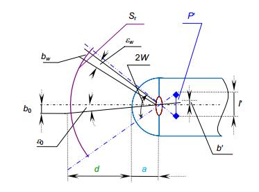
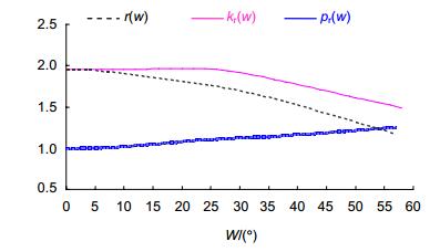
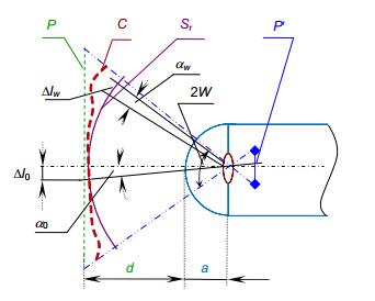
Overview: Based on clinical application, the capsule endoscope is designed as a wide field of view. However, the wide field of view will cause distortion. The distortion is easy to generate the matching inconsistency between the pixel element and the optical resolution element in the full field of view. Then the effectiveness of the pixels has declined. Furthermore, the distortion even causes the mismatching in some positions. The small intestine capsule endoscope is used to observe the surrounding field of view. The detection found that Moiré pattern occurred in the small intestine capsule endoscopy. Therefore, researches were carried out to establish the evaluation for the matching level between the optical resolution and the pixel number on the full field of view of the capsule endoscope, and the evaluation methods for the resolution validity of pixel number. This method selects the horizontal optical axis section as the meridian plane to analyze. The section passing through the planar array sensor is row or column scanned. The analytical method of the optical resolution angle continuation arrangement was adopted. Then the algorithm of line resolution element number was derived. The number of effective pixels of the line was described by the number of resolution elements. Then, the effective rate of line pixel number was obtained. The pixel element projection angle and the optical resolution angle were taken as the analytical units. The ratio between the pixel element projection angle and the optical resolution angle in the same field of view position was regarded as the line-matching ratio. Three basic functions for analysis were further extended, including the resolution angle density function, the relative function of line matching ratio and the density eigenfunction of pixel element projection angle. A simple measurement method was derived by using the simple conversion relations between a line segment and an included angle on a spherical field of view. Those three basic functions for analysis were obtained by fitting, so that the situation of the pixel element and the optical resolution element in every position, the matching details between two elements and the matching level between two elements in the full field of view were obtained. The parameters, including the line resolution element number, effective rate of line pixel number, center field of view matching ratio, maximum matching ratio of the full field of view and its position and other parameters, not only constitute the evaluation basis for matching ratio and resolution validity of the pixel number of the capsule endoscope, but also offer a reference for product design, analysis and modification.
-

-
表 1 测试及测算数据
Table 1. Test and calculate data
wj 0° 15° 25° 35° 45° Manufacturer A 's product Δlwj 71.3 71.3 71.3 75.5 82.4 mwj/m0 1 0.950 0.900 0.825 0.725 Manufacturer B 's product Δlwj 40.0 40.0 40.0 42.4 47.6 mwj/m0 1 0.966 0.897 0.793 0.621  下载: 导出CSV
表 2 测试结果
Table 2. Test result
参数 n0 n0/n k0 kw, m wm Manufacturer A 's product 211.5 0.44 0.4 0.5 57.5 Manufacturer B 's product 367.1 0.76 0.66 1.04 56.4
下载: 导出CSV
-
参考文献
[1] 数码相机中的图像传感器和信号处理[M].徐江涛, 高静, 聂海明, 译.北京: 清华大学出版社, 2015.
Nakamura J. Image Sensors and Signal Processing for Digital Still Cameras[M]. Xu J T, Gao J, Nie H M, trans. Beijing: Tsinghua University Press, 2015. Nakamura J.
[2] 王庆有.图像传感器应用技术[M]. 2版.北京:电子工业出版社, 2013.
[3] 陈远.基于垂直层叠结构的CMOS像素和图像传感器研究[D].杭州: 浙江大学, 2009.
Chen Y. Study of CMOS vertical integrated pixel and image sensor[D]. Hangzhou: Zhejiang University, 2009.
[4] 贾晓航, 张沁园, 段晓东, 等.医用内窥镜胶囊式内窥镜: YY 1298-2016[S].北京: 中国标准出版社, 2018.
Jia X H, Zhang Q Y, Duan X D, et al. Medical endoscopes—Capsule endoscopes: YY 1298-2016[S]. Beijing: China Standard Press, 2018.
[5] 贾晓航, 何涛, 颜青来, 等.医用内窥镜硬性内窥镜第1部分: 光学性能及测试方法: YY 0068.1-2008[S].北京: 中国标准出版社, 2009.
Jia X H, He T, Yan Q L, et al. Medical endoscopes—rigid endoscope—Part 1: optical properties and test methods: YY 0068.1-2008[S]. Beijing: China Standard Press, 2009.
[6] 贾晓航, 颜青来, 文燕.医用硬性内窥镜畸变的评定基础和方法[J].光学学报, 2006, 26(8): 1226-1230. doi: 10.3321/j.issn:0253-2239.2006.08.021
Jia X H, Yan Q L, Wen Y. Evaluation base and method of medical rigid endoscope distortion[J]. Acta Optica Sinica, 2006, 26(8): 1226-1230. doi: 10.3321/j.issn:0253-2239.2006.08.021
[7] 刘茂超, 张雷, 刘沛沛, 等. 300万像素手机镜头设计[J].应用光学, 2008, 29(6): 944-948. doi: 10.3969/j.issn.1002-2082.2008.06.023
Liu M C, Zhang L, Liu P P, et al. Design of lens for 3 mega-pixel mobile phone camera[J]. Journal of Applied Optics, 2008, 29(6): 944-948. doi: 10.3969/j.issn.1002-2082.2008.06.023
[8] 谷俊达, 向阳.电子内窥镜光学系统设计[J].长春理工大学学报(自然科学版), 2015, 38(2): 18-20, 24. doi: 10.3969/j.issn.1672-9870.2015.02.005
Gu J D, Xiang Y. Optical system design of electronic endoscope[J]. Journal of Changchun University of Science and Technology (Natural Science Edition), 2015, 38(2): 18-20, 24. doi: 10.3969/j.issn.1672-9870.2015.02.005
[9] 王军华, 卢景红, 徐敏.含有谐衍射面的单片式胶囊内窥镜光学设计[J].激光与光电子学进展, 2012, 49(12): 122203. 10.3788/lop49.122203
Wang J H, Lu J H, Xu M. Optical design of the single-chip capsule endoscopt with harmonic diffraction surface[J]. Laser & Optoelectronics Progress, 2012, 49(12): 122203. 10.3788/lop49.122203
[10] 朱佳巍, 丁桂林.一款超小型广角医用内窥镜镜头的设计[J].激光与光电子学进展, 2014, 51(9): 181-186.
Zhu J W, Ding G L. Design of an ultra-small wide-angle medical endoscope lens[J]. Laser & Optoelectronics Progress, 2014, 51(9): 181-186.
[11] 张艳辉, 黄战华.胶囊内窥镜技术的研究进展[J].现代仪器, 2006, 12(4): 4-7. doi: 10.3969/j.issn.1672-7916.2006.04.002
Zhang Y H, Huang Z H. The progress of capsule endoscope technology[J]. Modern Instruments, 2006, 12(4): 4-7. doi: 10.3969/j.issn.1672-7916.2006.04.002
[12] 吕联荣, 姜道连, 田春苗.电视原理及其应用技术[M].天津:天津大学出版社, 2001.
[13] 郭鑫, 张薇, 速晋辉, 等.可调焦胶囊内窥镜光学系统设计[J].光子学报, 2015, 44(5): 0522004. 10.3788/gzxb20154405.0522004
Guo X, Zhang W, Su J H, et al. Design of a focus-tunable capsule endoscope system[J]. Acta Photonica Sinica, 2015, 44(5): 0522004. 10.3788/gzxb20154405.0522004
[14] 叶斌, 王立强, 石岩, 等.高清医用电子内窥镜微型CMOS摄像模组(英文)[J].光子学报, 2010, 39(11): 1951-1955. 10.3788/gzxb20103911.1956
Ye B, Wang L Q, Shi Y, et al. High resolution miniaturized CMOS camera module for medical electronic endoscope[J]. Acta Photonica Sinica, 2010, 39(11): 1951-1955. 10.3788/gzxb20103911.1956
[15] 丁琴, 王惠南.采用GRIN透镜的微胶囊内窥成像技术[J].光子学报, 2004, 33(7): 889-892. 10.3969/j.issn.1000-3835.2011.02.014
Ding Q, Wang H N. An imaging technique of microcapsule endoscope using a GRIN lens[J]. Acta Photonica Sinica, 2004, 33(7): 889-892. 10.3969/j.issn.1000-3835.2011.02.014
[16] Tseng Y C, Han P, Hsu H C, et al. A flexible FOV capsule endoscope design based on compound lens[J]. Journal of Display Technology, 2016, 12(12): 1-7. doi: 10.1109/JDT.2017.2661078
[17] Ouyang M, Jeng W D. Design and analysis of radial imaging capsule endoscope (RICE) system[J]. Optics Express, 2011, 19(5): 4369-4383. doi: 10.1364/OE.19.004369
-
访问统计


 点击扫一扫
点击扫一扫
图(5)
表(2)
计量
- 文章访问数:
- PDF下载数:
- 施引文献: 0


